- PDB-2a4j: Solution structure of the C-terminal domain (T94-Y172) of the hum... -
+
Open data
ID or keywords:
Loading...
-
Basic information
Entry
Database: PDB / ID: 2a4j
Title
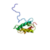
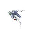
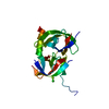
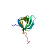
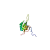
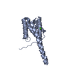
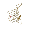
Solution structure of the C-terminal domain (T94-Y172) of the human centrin 2 in complex with a 17 residues peptide (P1-XPC) from xeroderma pigmentosum group C protein
Components
17-mer peptide P1-XPC from DNA-repair protein complementing XP-C cells
Centrin 2
Keywords
STRUCTURAL PROTEIN / EF-hand
Function / homology
Function and homology information
heteroduplex DNA loop binding / nucleotide-excision repair factor 2 complex / pyrimidine dimer repair by nucleotide-excision repair / XPC complex / 9+2 motile cilium / nucleotide-excision repair complex / photoreceptor connecting cilium / DNA damage sensor activity / heterotrimeric G-protein binding / transcription export complex 2 ...heteroduplex DNA loop binding / nucleotide-excision repair factor 2 complex / pyrimidine dimer repair by nucleotide-excision repair / XPC complex / 9+2 motile cilium / nucleotide-excision repair complex / photoreceptor connecting cilium / DNA damage sensor activity / heterotrimeric G-protein binding / transcription export complex 2 / nuclear pore nuclear basket / response to auditory stimulus / bubble DNA binding / response to UV-B / mitotic intra-S DNA damage checkpoint signaling / UV-damage excision repair / regulation of mitotic cell cycle phase transition / centriole replication / glial cell projection / mRNA transport / mismatch repair / SUMOylation of DNA damage response and repair proteins / site of DNA damage / Loss of Nlp from mitotic centrosomes / Loss of proteins required for interphase microtubule organization from the centrosome / Recruitment of mitotic centrosome proteins and complexes / Recruitment of NuMA to mitotic centrosomes / Anchoring of the basal body to the plasma membrane / AURKA Activation by TPX2 / regulation of cytokinesis / nucleotide-excision repair / centriole / microtubule cytoskeleton organization / DNA Damage Recognition in GG-NER / G-protein beta/gamma-subunit complex binding / apical part of cell / Formation of Incision Complex in GG-NER / Regulation of PLK1 Activity at G2/M Transition / mitotic cell cycle / protein transport / single-stranded DNA binding / spermatogenesis / microtubule binding / damaged DNA binding / RNA polymerase II-specific DNA-binding transcription factor binding / transcription coactivator activity / ciliary basal body / response to xenobiotic stimulus / cell division / DNA repair / calcium ion binding / centrosome / positive regulation of DNA-templated transcription / chromatin / protein-containing complex binding / nucleolus / mitochondrion / nucleoplasm / nucleus / plasma membrane / cytoplasm / cytosol Similarity search - Function
Method: simulated annealing / Software ordinal: 1 Details: the structures are based on 1298 NOE distance restraints, 121 dihedral angle restraints, and 84 hydrogen bond restraints.
NMR representative
Selection criteria: fewest violations
NMR ensemble
Conformer selection criteria: structures with the least restraint violations Conformers calculated total number: 200 / Conformers submitted total number: 20
+
About Yorodumi
-
News
-
Feb 9, 2022. New format data for meta-information of EMDB entries
New format data for meta-information of EMDB entries
Version 3 of the EMDB header file is now the official format.
The previous official version 1.9 will be removed from the archive.
In the structure databanks used in Yorodumi, some data are registered as the other names, "COVID-19 virus" and "2019-nCoV". Here are the details of the virus and the list of structure data.
Jan 31, 2019. EMDB accession codes are about to change! (news from PDBe EMDB page)
EMDB accession codes are about to change! (news from PDBe EMDB page)
The allocation of 4 digits for EMDB accession codes will soon come to an end. Whilst these codes will remain in use, new EMDB accession codes will include an additional digit and will expand incrementally as the available range of codes is exhausted. The current 4-digit format prefixed with “EMD-” (i.e. EMD-XXXX) will advance to a 5-digit format (i.e. EMD-XXXXX), and so on. It is currently estimated that the 4-digit codes will be depleted around Spring 2019, at which point the 5-digit format will come into force.
The EM Navigator/Yorodumi systems omit the EMD- prefix.
Related info.:Q: What is EMD? / ID/Accession-code notation in Yorodumi/EM Navigator
Yorodumi is a browser for structure data from EMDB, PDB, SASBDB, etc.
This page is also the successor to EM Navigator detail page, and also detail information page/front-end page for Omokage search.
The word "yorodu" (or yorozu) is an old Japanese word meaning "ten thousand". "mi" (miru) is to see.
Related info.:EMDB / PDB / SASBDB / Comparison of 3 databanks / Yorodumi Search / Aug 31, 2016. New EM Navigator & Yorodumi / Yorodumi Papers / Jmol/JSmol / Function and homology information / Changes in new EM Navigator and Yorodumi
 Movie
Movie Controller

 Yorodumi
 Open data
 Basic information
 Components
 Keywords
 Function and homology information
 Homo sapiens (human)
 Authors
 Citation
 Structure visualization
 Downloads & links
 Other downloads
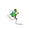
 PDBj
PDBj


 Assembly

 HSQC
HSQC Sample preparation
 Processing