- PDB-2a2a: High-resolution crystallographic analysis of the autoinhibited co... -
+
Open data
ID or keywords:
Loading...
-
Basic information
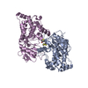
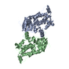
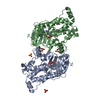
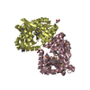
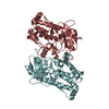
Entry
Database: PDB / ID: 2a2a
Title
High-resolution crystallographic analysis of the autoinhibited conformation of a human death-associated protein kinase
Components
Death-associated protein kinase 2
Keywords
TRANSFERASE / protein kinase / autoinhibition
Function / homology
Function and homology information
positive regulation of eosinophil chemotaxis / autophagosome lumen / regulation of intrinsic apoptotic signaling pathway / neutrophil migration / Caspase activation via Dependence Receptors in the absence of ligand / anoikis / positive regulation of neutrophil chemotaxis / regulation of autophagy / protein autophosphorylation / cytoplasmic vesicle ...positive regulation of eosinophil chemotaxis / autophagosome lumen / regulation of intrinsic apoptotic signaling pathway / neutrophil migration / Caspase activation via Dependence Receptors in the absence of ligand / anoikis / positive regulation of neutrophil chemotaxis / regulation of autophagy / protein autophosphorylation / cytoplasmic vesicle / regulation of apoptotic process / protein phosphorylation / calmodulin binding / non-specific serine/threonine protein kinase / intracellular signal transduction / positive regulation of apoptotic process / protein serine kinase activity / protein serine/threonine kinase activity / apoptotic process / Golgi apparatus / ATP binding / identical protein binding / nucleus / cytoplasm Similarity search - Function
Phosphorylase Kinase; domain 1 / Phosphorylase Kinase; domain 1 / Transferase(Phosphotransferase) domain 1 / Transferase(Phosphotransferase); domain 1 / Serine/threonine-protein kinase, active site / Serine/Threonine protein kinases active-site signature. / Protein kinase domain / Serine/Threonine protein kinases, catalytic domain / Protein kinase, ATP binding site / Protein kinases ATP-binding region signature. ...Phosphorylase Kinase; domain 1 / Phosphorylase Kinase; domain 1 / Transferase(Phosphotransferase) domain 1 / Transferase(Phosphotransferase); domain 1 / Serine/threonine-protein kinase, active site / Serine/Threonine protein kinases active-site signature. / Protein kinase domain / Serine/Threonine protein kinases, catalytic domain / Protein kinase, ATP binding site / Protein kinases ATP-binding region signature. / Protein kinase domain profile. / Protein kinase domain / Protein kinase-like domain superfamily / 2-Layer Sandwich / Orthogonal Bundle / Mainly Alpha / Alpha Beta Similarity search - Domain/homology
Mass: 18.015 Da / Num. of mol.: 1371 / Source method: isolated from a natural source / Formula: H2O
-
Details
Sequence details
There are differences between the seqres and the sequence database. The depositors say that there ...There are differences between the seqres and the sequence database. The depositors say that there are two versions of the sequence in the databases and the sequence in this protein corresponds to the other one and is thus correct. The difference is evidenced in the REFERENCE: JOURNAL Mol. Cell. Biol. 20 (3), 1044-1054 (2000) PUBMED 10629061
-
Experimental details
-
Experiment
Experiment
Method: X-RAY DIFFRACTION / Number of used crystals: 1
-
Sample preparation
Crystal
Density Matthews: 2.2 Å3/Da / Density % sol: 43 %
Crystal grow
Temperature: 295 K / Method: vapor diffusion, sitting drop / pH: 8.5 Details: PEG4000, glycerol, DTT, Tris, lithium sulfate, pH 8.5, VAPOR DIFFUSION, SITTING DROP, temperature 295K
In the structure databanks used in Yorodumi, some data are registered as the other names, "COVID-19 virus" and "2019-nCoV". Here are the details of the virus and the list of structure data.
Jan 31, 2019. EMDB accession codes are about to change! (news from PDBe EMDB page)
EMDB accession codes are about to change! (news from PDBe EMDB page)
The allocation of 4 digits for EMDB accession codes will soon come to an end. Whilst these codes will remain in use, new EMDB accession codes will include an additional digit and will expand incrementally as the available range of codes is exhausted. The current 4-digit format prefixed with “EMD-” (i.e. EMD-XXXX) will advance to a 5-digit format (i.e. EMD-XXXXX), and so on. It is currently estimated that the 4-digit codes will be depleted around Spring 2019, at which point the 5-digit format will come into force.
The EM Navigator/Yorodumi systems omit the EMD- prefix.
Related info.:Q: What is EMD? / ID/Accession-code notation in Yorodumi/EM Navigator
Yorodumi is a browser for structure data from EMDB, PDB, SASBDB, etc.
This page is also the successor to EM Navigator detail page, and also detail information page/front-end page for Omokage search.
The word "yorodu" (or yorozu) is an old Japanese word meaning "ten thousand". "mi" (miru) is to see.
Related info.:EMDB / PDB / SASBDB / Comparison of 3 databanks / Yorodumi Search / Aug 31, 2016. New EM Navigator & Yorodumi / Yorodumi Papers / Jmol/JSmol / Function and homology information / Changes in new EM Navigator and Yorodumi
 Movie
Movie Controller

 Yorodumi
 Open data
 Basic information
 Components
 Keywords
 Function and homology information
 Homo sapiens (human)
 X-RAY DIFFRACTION /
  Authors
 Citation
 Structure visualization
 Downloads & links
 Other downloads


 PDBj
PDBj


 Assembly


 Mass: 22.990 Da / Num. of mol.: 8 / Source method: obtained synthetically / Formula: Na
 Mass: 35.453 Da / Num. of mol.: 3 / Source method: obtained synthetically / Formula: Cl
 Mass: 154.251 Da / Num. of mol.: 4 / Source method: obtained synthetically / Formula: C4H10O2S2
 Mass: 92.094 Da / Num. of mol.: 3 / Source method: obtained synthetically / Formula: C3H8O3
 Sample preparation
 / Beamline: BW7A / Wavelength: 0.9206 Å
 Processing