 Movie
Movie Controller

[English] 日本語
 Yorodumi
- PDB-2a28: Atomic-resolution crystal structure of the second SH3 domain of y... -
+ Open data

- Basic information
| Entry | Database: PDB / ID: 2a28 | ||||||
|---|---|---|---|---|---|---|---|
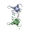
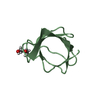
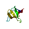
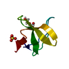
| Title | Atomic-resolution crystal structure of the second SH3 domain of yeast Bzz1 determined from a pseudomerohedrally twinned crystal | ||||||
 Components Components | BZZ1 protein | ||||||
 Keywords Keywords | SIGNALING PROTEIN / SH3 domain | ||||||
| Function / homology |  Function and homology information Function and homology informationactin nucleation / actin cortical patch / regulation of actin filament polymerization / cellular bud neck / mating projection tip / response to salt stress / actin filament organization / phospholipid binding / enzyme activator activity / endocytosis ...actin nucleation / actin cortical patch / regulation of actin filament polymerization / cellular bud neck / mating projection tip / response to salt stress / actin filament organization / phospholipid binding / enzyme activator activity / endocytosis / plasma membrane / cytoplasm Similarity search - Function | ||||||
| Biological species |  | ||||||
| Method |  X-RAY DIFFRACTION / SYNCHROTRON / MOLECULAR REPLACEMENT / Resolution: 1.07 Å X-RAY DIFFRACTION / SYNCHROTRON / MOLECULAR REPLACEMENT / Resolution: 1.07 Å | ||||||
 Authors Authors | Kursula, P. / Kursula, I. / Lehmann, F. / Zou, P. / Song, Y.H. / Wilmanns, M. | ||||||
 Citation Citation | Journal: To be Published Title: Structural genomics of yeast SH3 domains Authors: Kursula, P. / Kursula, I. / Lehmann, F. / Zou, P. / Song, Y.H. / Wilmanns, M. | ||||||
| History |
|
- Structure visualization
| Structure viewer | Molecule: MolmilJmol/JSmol |
|---|
- Downloads & links
-Download
| PDBx/mmCIF format | 2a28.cif.gz | 107.9 KB | Display | PDBx/mmCIF format |
|---|---|---|---|---|
| PDB format | pdb2a28.ent.gz | 84 KB | Display | PDB format |
| PDBx/mmJSON format | 2a28.json.gz | Tree view | PDBx/mmJSON format | |
| Others |  Other downloads Other downloads |
-Validation report
| Arichive directory | https://data.pdbj.org/pub/pdb/validation_reports/a2/2a28ftp://data.pdbj.org/pub/pdb/validation_reports/a2/2a28 | HTTPS FTP |
|---|
-Related structure data


-Links
 PDBj
PDBj
- Assembly
| Deposited unit | 
| ||||||||
|---|---|---|---|---|---|---|---|---|---|
| 1 | 
| ||||||||
| 2 | 
| ||||||||
| 3 | 
| ||||||||
| 4 | 
| ||||||||
| Unit cell |
|
-Components
| #1: Protein | Mass: 5768.271 Da / Num. of mol.: 4 / Fragment: SH3 domain Source method: isolated from a genetically manipulated source Source: (gene. exp.) Plasmid: pDEST-17 / Production host:  #2: Water | ChemComp-HOH / |  Mass: 18.015 Da / Num. of mol.: 390 / Source method: isolated from a natural source / Formula: H2O Mass: 18.015 Da / Num. of mol.: 390 / Source method: isolated from a natural source / Formula: H2O |
|---|
-Experimental details
-Experiment
| Experiment | Method: X-RAY DIFFRACTION / Number of used crystals: 1 |
|---|
- Sample preparation
| Crystal | Density Matthews: 2.2 Å3/Da / Density % sol: 44 % |
|---|---|
| Crystal grow | Temperature: 295 K / Method: vapor diffusion, hanging drop / pH: 8.5 Details: ammonium sulfate, pH 8.5, VAPOR DIFFUSION, HANGING DROP, temperature 295K |
-Data collection
| Diffraction | Mean temperature: 100 K |
|---|---|
| Diffraction source | Source: SYNCHROTRON / Site: EMBL/DESY, HAMBURG  / Beamline: BW7A / Wavelength: 0.8126 Å / Beamline: BW7A / Wavelength: 0.8126 Å |
| Detector | Type: MARRESEARCH / Detector: CCD / Date: Mar 7, 2005 |
| Radiation | Protocol: SINGLE WAVELENGTH / Monochromatic (M) / Laue (L): M / Scattering type: x-ray |
| Radiation wavelength | Wavelength: 0.8126 Å / Relative weight: 1 |
| Reflection | Resolution: 1.07→30 Å / Num. all: 84816 / Num. obs: 84816 / % possible obs: 96 % / Observed criterion σ(F): -3 / Observed criterion σ(I): -3 / Redundancy: 3.2 % / Biso Wilson estimate: 11.9 Å2 / Rsym value: 0.045 / Net I/σ(I): 16.2 |
| Reflection shell | Resolution: 1.07→1.1 Å / Redundancy: 2.4 % / Mean I/σ(I) obs: 4.1 / Num. unique all: 6490 / Rsym value: 0.241 / % possible all: 93.1 |
- Processing
| Software |
| |||||||||||||||||||||||||||||||||
|---|---|---|---|---|---|---|---|---|---|---|---|---|---|---|---|---|---|---|---|---|---|---|---|---|---|---|---|---|---|---|---|---|---|---|
| Refinement | Method to determine structure: MOLECULAR REPLACEMENT / Resolution: 1.07→20 Å / Num. parameters: 18238 / Num. restraintsaints: 21490 / Isotropic thermal model: ANISOTROPIC REFINEMENT / Cross valid method: FREE R / σ(F): 0 / Stereochemistry target values: Engh & Huber Details: The crystal form is twinned by the operator h,-k,-l, twinning fraction 37 %.
| |||||||||||||||||||||||||||||||||
| Refine analyze | Num. disordered residues: 4 / Occupancy sum hydrogen: 1428 / Occupancy sum non hydrogen: 2007 | |||||||||||||||||||||||||||||||||
| Refinement step | Cycle: LAST / Resolution: 1.07→20 Å
| |||||||||||||||||||||||||||||||||
| Refine LS restraints |
|
