Movie
 Controller


[English] 日本語
 Yorodumi
- PDB-29ui: FAM72A-UNG2 bound to the discoidin domain of substrate receptor MKLN1 -
+ Open data

- Basic information
| Entry | Database: PDB / ID: 29ui | |||||||||
|---|---|---|---|---|---|---|---|---|---|---|
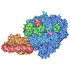
| Title | FAM72A-UNG2 bound to the discoidin domain of substrate receptor MKLN1 | |||||||||
 Components |
| |||||||||
 Keywords | LIGASE / complex | |||||||||
| Function / homology |  Function and homology information postsynaptic specialization membrane of symmetric synapse / postsynaptic endosome membrane / neurotransmitter receptor transport postsynaptic membrane to endosome / GID complex / base-excision repair, AP site formation via deaminated base removal / regulation of receptor internalization / uracil-DNA glycosylase / depyrimidination / Displacement of DNA glycosylase by APEX1 / uracil DNA N-glycosylase activity ...postsynaptic specialization membrane of symmetric synapse / postsynaptic endosome membrane / neurotransmitter receptor transport postsynaptic membrane to endosome / GID complex / base-excision repair, AP site formation via deaminated base removal / regulation of receptor internalization / uracil-DNA glycosylase / depyrimidination / Displacement of DNA glycosylase by APEX1 / uracil DNA N-glycosylase activity / single strand break repair / ribosomal small subunit binding / cell-matrix adhesion / ruffle / ubiquitin ligase complex / Recognition and association of DNA glycosylase with site containing an affected pyrimidine / Cleavage of the damaged pyrimidine / Regulation of pyruvate metabolism / Chromatin modifications during the maternal to zygotic transition (MZT) / regulation of cell shape / GABA-ergic synapse / base-excision repair / actin cytoskeleton organization / cell cortex / proteasome-mediated ubiquitin-dependent protein catabolic process / damaged DNA binding / postsynapse / negative regulation of apoptotic process / synapse / signal transduction / protein homodimerization activity / mitochondrion / nucleoplasm / identical protein binding / nucleus / cytosol / cytoplasm Similarity search - Function | |||||||||
| Biological species |  Homo sapiens (human) | |||||||||
| Method | ELECTRON MICROSCOPY / single particle reconstruction / cryo EM / Resolution: 3 Å | |||||||||
 Authors | Sepic, S. / Du, J. / Prabu, J.R. / Schulman, B.A. | |||||||||
| Funding support |  Germany, 1items
| |||||||||
 Citation | Journal: Nat Commun / Year: 2026 Title: Multimodal substrate recruitment enables CTLH E3 ligase to target acetylated N-, C-, and internal degrons. Authors: Sara Sepic / Karthik V Gottemukkala / Jiale Du / Samuel A Maiwald / Jakub Chrustowicz / Eleftheria C Papadopoulou / Luca J Stier / Susanne von Gronau / Brenda A Schulman / Abstract: The GID/CTLH family of E3 ubiquitin ligases employs several substrate receptor subunits that recruit distinct degrons, but the substrate recognition mechanisms of the CTLH assembly have largely ...The GID/CTLH family of E3 ubiquitin ligases employs several substrate receptor subunits that recruit distinct degrons, but the substrate recognition mechanisms of the CTLH assembly have largely remained elusive. Here, we reconstitute CTLH-dependent ubiquitylation of three biochemically distinct substrates - MKLN1 itself, ZMYND19, and FAM72A-recruited UNG2, and determine cryo-EM structures of each substrate bound to MKLN1. The structures reveal how MKLN1's discoidin and Kelch β-propeller domains engage substrates through multivalent contacts. MKLN1 recruits itself through discoidin-Kelch interactions between MKLN1 dimers, forming an assembly that competes with other substrates. ZMYND19 and FAM72A each bind to MKLN1 through loops engaging the discoidin trench domain while their C-terminal Arg residues engage the MKLN1 Kelch central channel, identifying MKLN1 as an Arg/C-degron receptor. The acetylated N-terminus of UNG2 is positioned within a Y-shaped tunnel of FAM72A, defining an adaptor-mediated Ac/N-degron recognition mechanism. Thus, our data reveal how combinatorial deployment of MKLN1 discoidin and Kelch domains enable a single receptor subunit to recognize a diverse substrate repertoire, adding to a complex picture of degron recognition across the GID/CTLH family. | |||||||||
| History |
|
- Structure visualization
| Structure viewer | Molecule: MolmilJmol/JSmol |
|---|
- Downloads & links
-Download
| PDBx/mmCIF format | 29ui.cif.gz | 202.2 KB | Display | PDBx/mmCIF format |
|---|---|---|---|---|
| PDB format | pdb29ui.ent.gz | 144.6 KB | Display | PDB format |
| PDBx/mmJSON format | 29ui.json.gz | Tree view | PDBx/mmJSON format | |
| Others |  Other downloads |
-Validation report
| Arichive directory | https://data.pdbj.org/pub/pdb/validation_reports/9u/29uiftp://data.pdbj.org/pub/pdb/validation_reports/9u/29ui | HTTPS FTP |
|---|
-Related structure data
| Related structure data |  57387MC  29ueC  29ugC  31tmC M: map data used to model this data C: citing same article ( |
|---|---|
| Similar structure data |


-Links
 PDBj
- Assembly
| Deposited unit | 
|
|---|---|
| 1 |
|
-Components
| #1: Protein | Mass: 16636.326 Da / Num. of mol.: 2 Source method: isolated from a genetically manipulated source Source: (gene. exp.) Homo sapiens (human) / Gene: FAM72A, UGENE / Production host:  Trichoplusia ni (cabbage looper) / References: UniProt: Q5TYM5 #2: Protein | Mass: 34722.473 Da / Num. of mol.: 2 Source method: isolated from a genetically manipulated source Source: (gene. exp.) Homo sapiens (human) / Gene: UNG, DGU, UNG1, UNG15 / Production host: Trichoplusia ni (cabbage looper) / References: UniProt: P13051, uracil-DNA glycosylase#3: Protein | Mass: 84663.484 Da / Num. of mol.: 2 Source method: isolated from a genetically manipulated source Source: (gene. exp.) Homo sapiens (human) / Gene: MKLN1 / Production host: Trichoplusia ni (cabbage looper) / References: UniProt: Q9UL63#4: Chemical | ChemComp-ZN /   Mass: 65.409 Da / Num. of mol.: 4 / Source method: obtained synthetically / Formula: Zn / Feature type: SUBJECT OF INVESTIGATION Has ligand of interest | Y | Has protein modification | Y | |
|---|
-Experimental details
-Experiment
| Experiment | Method: ELECTRON MICROSCOPY |
|---|---|
| EM experiment | Aggregation state: PARTICLE / 3D reconstruction method: single particle reconstruction |
- Sample preparation
| Component | Name: Structure of FAM72A-UNG2 bound to MKLN1 discoidin domain Type: COMPLEX / Entity ID: #1-#3 / Source: RECOMBINANT |
|---|---|
| Source (natural) | Organism: Homo sapiens (human) |
| Source (recombinant) | Organism: Trichoplusia ni (cabbage looper) |
| Buffer solution | pH: 7.5 |
| Specimen | Embedding applied: NO / Shadowing applied: NO / Staining applied: NO / Vitrification applied: YES |
| Vitrification | Cryogen name: ETHANE-PROPANE |
- Electron microscopy imaging
| Experimental equipment | 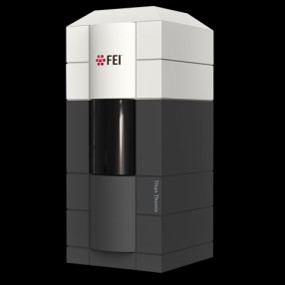 Model: Titan Krios / Image courtesy: FEI Company |
|---|---|
| Microscopy | Model: TFS KRIOS |
| Electron gun | Electron source:  FIELD EMISSION GUN / Accelerating voltage: 300 kV / Illumination mode: FLOOD BEAM |
| Electron lens | Mode: BRIGHT FIELD / Nominal defocus max: 2600 nm / Nominal defocus min: 1000 nm |
| Specimen holder | Cryogen: NITROGEN |
| Image recording | Electron dose: 60 e/Å2 / Film or detector model: GATAN K3 BIOQUANTUM (6k x 4k) |
- Processing
| EM software |
| ||||||||||||||||||||||||
|---|---|---|---|---|---|---|---|---|---|---|---|---|---|---|---|---|---|---|---|---|---|---|---|---|---|
| CTF correction | Type: PHASE FLIPPING AND AMPLITUDE CORRECTION | ||||||||||||||||||||||||
| 3D reconstruction | Resolution: 3 Å / Resolution method: FSC 0.143 CUT-OFF / Num. of particles: 1117642 / Symmetry type: POINT | ||||||||||||||||||||||||
| Refinement | Highest resolution: 3 Å Stereochemistry target values: REAL-SPACE (WEIGHTED MAP SUM AT ATOM CENTERS) | ||||||||||||||||||||||||
| Refine LS restraints |
|