 Movie
Movie Controller

[English] 日本語
 Yorodumi
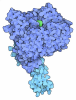
- PDB-28mj: Cryo-EM structure of UBA6 in complex with FAT10 in the pre-adenyl... -
+ Open data

- Basic information
| Entry | Database: PDB / ID: 28mj | |||||||||
|---|---|---|---|---|---|---|---|---|---|---|
| Title | Cryo-EM structure of UBA6 in complex with FAT10 in the pre-adenylation state. | |||||||||
 Components Components |
| |||||||||
 Keywords Keywords | SIGNALING PROTEIN / Ubiquitin-activating enzyme / ubiquitin-like protein / adenylation / signalling cascade | |||||||||
| Function / homology |  Function and homology information Function and homology informationprotein modification by small protein conjugation / FAT10 activating enzyme activity / aggresome assembly / myeloid dendritic cell differentiation / E1 ubiquitin-activating enzyme / ubiquitin activating enzyme activity / aggresome / regulation of mitotic cell cycle phase transition / proteasome binding / response to tumor necrosis factor ...protein modification by small protein conjugation / FAT10 activating enzyme activity / aggresome assembly / myeloid dendritic cell differentiation / E1 ubiquitin-activating enzyme / ubiquitin activating enzyme activity / aggresome / regulation of mitotic cell cycle phase transition / proteasome binding / response to tumor necrosis factor / Dengue Virus Attachment and Entry / response to type II interferon / Synthesis of active ubiquitin: roles of E1 and E2 enzymes / Antigen processing: Ubiquitination & Proteasome degradation / Neddylation / ubiquitin-dependent protein catabolic process / positive regulation of canonical NF-kappaB signal transduction / protein ubiquitination / positive regulation of apoptotic process / DNA damage response / proteolysis / nucleoplasm / metal ion binding / ATP binding / nucleus / cytosol / cytoplasm Similarity search - Function | |||||||||
| Biological species |  Homo sapiens (human) Homo sapiens (human) | |||||||||
| Method | ELECTRON MICROSCOPY / single particle reconstruction / cryo EM / Resolution: 2.77 Å | |||||||||
 Authors Authors | Ellison, C.J. / Dalietou, E.V. / Riechmann, C. / Elliott, P.R. | |||||||||
| Funding support |  United Kingdom, 2items United Kingdom, 2items
| |||||||||
 Citation Citation | Journal: Nat Commun / Year: 2026 Title: Structural determinants for FAT10 activation and transfer from UBA6 to E2 enzymes. Authors: Cara J Ellison / Carlos Riechmann / Evmorfia V Dalietou / Michael D R Simmons / Emma C Dodd / Paul R Elliott / Abstract: Attachment of the ubiquitin-like protein (UBL) FAT10 onto substrates targets them for proteasomal degradation. Like ubiquitin, FAT10 is activated by the E1 enzyme UBA6 then transferred to E2 enzymes, ...Attachment of the ubiquitin-like protein (UBL) FAT10 onto substrates targets them for proteasomal degradation. Like ubiquitin, FAT10 is activated by the E1 enzyme UBA6 then transferred to E2 enzymes, but mechanisms controlling ubiquitin versus FAT10 activation by UBA6 and FAT10 transfer onto E2s remain unclear. Using cryo-EM, we visualise all stages of FAT10 E1-E2 handover: adenylation, thiolation and transthiolation. We find that FAT10 monopolises UBA6 by out-competing ubiquitin for thiolation and blocking the adenylation domain, preventing further UBL recruitment and promoting FAT10 signalling. We profiled UBA6-compatible E2 enzymes and found FAT10 transfer is restricted to a select subset associated with specific cellular pathways. UBE2Z (USE1) showed highest activity followed by UBE2D2, UBE2J2 and UBE2S. Capturing FAT10 or ubiquitin transfer from UBA6 to UBE2Z reveals UBE2Z is highly specialised for FAT10 transfer. It simultaneously engages both FAT10 domains (UBL1 and UBL2) and co-ordinates the metabolite inositol hexakisphosphate (InsP) bound within the UBA6 catalytic domain. This InsP co-ordination extends to other FAT10 compatible E2s. Together, our structural and biochemical analyses reveal regulatory mechanisms underpinning FAT10 activation and transfer. We define principles governing selective FAT10 transfer, highlighting favourable interactions with FAT10 C-terminal domain (UBL2) and stable UBA6 binding, ensuring controlled conjugation onto substrates. | |||||||||
| History |
|
- Structure visualization
| Structure viewer | Molecule: MolmilJmol/JSmol |
|---|
- Downloads & links
-Download
| PDBx/mmCIF format | 28mj.cif.gz | 565.2 KB | Display | PDBx/mmCIF format |
|---|---|---|---|---|
| PDB format | pdb28mj.ent.gz | 371.5 KB | Display | PDB format |
| PDBx/mmJSON format | 28mj.json.gz | Tree view | PDBx/mmJSON format | |
| Others |  Other downloads Other downloads |
-Validation report
| Arichive directory | https://data.pdbj.org/pub/pdb/validation_reports/8m/28mjftp://data.pdbj.org/pub/pdb/validation_reports/8m/28mj | HTTPS FTP |
|---|
-Related structure data
| Related structure data |  56614MC  28mkC  28moC  28mpC  28mqC M: map data used to model this data C: citing same article ( |
|---|---|
| Similar structure data |


-Links
 PDBj
PDBj

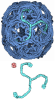

- Assembly
| Deposited unit | 
|
|---|---|
| 1 |
|
-Components
| #1: Protein | Mass: 18630.637 Da / Num. of mol.: 1 / Mutation: C7T, C9T, C134L, C160S, C162S Source method: isolated from a genetically manipulated source Source: (gene. exp.) Homo sapiens (human) / Gene: UBD, FAT10 / Production host:   Escherichia coli (E. coli) / References: UniProt: O15205 Escherichia coli (E. coli) / References: UniProt: O15205 |
|---|---|
| #2: Protein | Mass: 118266.883 Da / Num. of mol.: 1 Source method: isolated from a genetically manipulated source Source: (gene. exp.) Homo sapiens (human) / Gene: UBA6, MOP4, UBE1L2 / Production host:   Spodoptera frugiperda (fall armyworm) / References: UniProt: A0AVT1, E1 ubiquitin-activating enzyme Spodoptera frugiperda (fall armyworm) / References: UniProt: A0AVT1, E1 ubiquitin-activating enzyme |
| #3: Chemical | ChemComp-IHP / 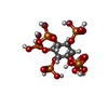 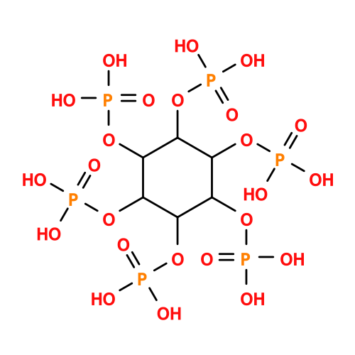 Mass: 660.035 Da / Num. of mol.: 1 / Source method: obtained synthetically / Formula: C6H18O24P6 / Feature type: SUBJECT OF INVESTIGATION Mass: 660.035 Da / Num. of mol.: 1 / Source method: obtained synthetically / Formula: C6H18O24P6 / Feature type: SUBJECT OF INVESTIGATION |
| #4: Chemical | ChemComp-ATP / 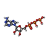 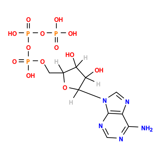 Mass: 507.181 Da / Num. of mol.: 1 / Source method: obtained synthetically / Formula: C10H16N5O13P3 / Feature type: SUBJECT OF INVESTIGATION / Comment: ATP, energy-carrying molecule*YM Mass: 507.181 Da / Num. of mol.: 1 / Source method: obtained synthetically / Formula: C10H16N5O13P3 / Feature type: SUBJECT OF INVESTIGATION / Comment: ATP, energy-carrying molecule*YM |
| #5: Chemical | ChemComp-MG /   Mass: 24.305 Da / Num. of mol.: 1 / Source method: obtained synthetically / Formula: Mg / Feature type: SUBJECT OF INVESTIGATION Mass: 24.305 Da / Num. of mol.: 1 / Source method: obtained synthetically / Formula: Mg / Feature type: SUBJECT OF INVESTIGATION |
| Has ligand of interest | Y |
| Has protein modification | N |
-Experimental details
-Experiment
| Experiment | Method: ELECTRON MICROSCOPY |
|---|---|
| EM experiment | Aggregation state: PARTICLE / 3D reconstruction method: single particle reconstruction |
- Sample preparation
| Component | Name: UBA6 in complex with FAT10 in the pre-adenylation state. Type: COMPLEX / Entity ID: #1-#2 / Source: RECOMBINANT |
|---|---|
| Source (natural) | Organism: Homo sapiens (human) |
| Source (recombinant) | Organism: Spodoptera frugiperda (fall armyworm) |
| Buffer solution | pH: 7.5 |
| Specimen | Embedding applied: NO / Shadowing applied: NO / Staining applied: NO / Vitrification applied: YES |
| Vitrification | Cryogen name: ETHANE |
- Electron microscopy imaging
| Experimental equipment |  Model: Titan Krios / Image courtesy: FEI Company |
|---|---|
| Microscopy | Model: TFS KRIOS |
| Electron gun | Electron source:  FIELD EMISSION GUN / Accelerating voltage: 300 kV / Illumination mode: FLOOD BEAM FIELD EMISSION GUN / Accelerating voltage: 300 kV / Illumination mode: FLOOD BEAM |
| Electron lens | Mode: BRIGHT FIELD / Nominal defocus max: 2400 nm / Nominal defocus min: 800 nm |
| Image recording | Electron dose: 42.2 e/Å2 / Film or detector model: GATAN K3 BIOQUANTUM (6k x 4k) |
- Processing
| EM software |
| ||||||||||||||||||||||||
|---|---|---|---|---|---|---|---|---|---|---|---|---|---|---|---|---|---|---|---|---|---|---|---|---|---|
| CTF correction | Type: PHASE FLIPPING AND AMPLITUDE CORRECTION | ||||||||||||||||||||||||
| 3D reconstruction | Resolution: 2.77 Å / Resolution method: FSC 0.143 CUT-OFF / Num. of particles: 795937 / Symmetry type: POINT | ||||||||||||||||||||||||
| Refinement | Cross valid method: NONE Stereochemistry target values: GeoStd + Monomer Library + CDL v1.2 | ||||||||||||||||||||||||
| Displacement parameters | Biso mean: 164.13 Å2 | ||||||||||||||||||||||||
| Refine LS restraints |
|
