 Movie
Movie Controller

[English] 日本語
 Yorodumi
- EMDB-56646: Cryo-EM structure of UBA6-FAT10Dha-UBE2Z trapped ternary complex -
+ Open data

- Basic information
| Entry |  | |||||||||
|---|---|---|---|---|---|---|---|---|---|---|
| Title | Cryo-EM structure of UBA6-FAT10Dha-UBE2Z trapped ternary complex | |||||||||
 Map data Map data | ||||||||||
 Sample Sample |
| |||||||||
 Keywords Keywords | Ubiquitin-activating enzyme / ubiquitin-like protein / adenylation / signalling cascade / SIGNALING PROTEIN | |||||||||
| Function / homology |  Function and homology information Function and homology informationprotein modification by small protein conjugation / FAT10 activating enzyme activity / aggresome assembly / myeloid dendritic cell differentiation / E1 ubiquitin-activating enzyme / ubiquitin activating enzyme activity / aggresome / E2 ubiquitin-conjugating enzyme / regulation of mitotic cell cycle phase transition / proteasome binding ...protein modification by small protein conjugation / FAT10 activating enzyme activity / aggresome assembly / myeloid dendritic cell differentiation / E1 ubiquitin-activating enzyme / ubiquitin activating enzyme activity / aggresome / E2 ubiquitin-conjugating enzyme / regulation of mitotic cell cycle phase transition / proteasome binding / ubiquitin conjugating enzyme activity / response to tumor necrosis factor / Dengue Virus Attachment and Entry / response to type II interferon / Synthesis of active ubiquitin: roles of E1 and E2 enzymes / Antigen processing: Ubiquitination & Proteasome degradation / Neddylation / ubiquitin-dependent protein catabolic process / molecular adaptor activity / positive regulation of canonical NF-kappaB signal transduction / protein ubiquitination / positive regulation of apoptotic process / apoptotic process / DNA damage response / negative regulation of apoptotic process / proteolysis / nucleoplasm / metal ion binding / ATP binding / nucleus / cytosol / cytoplasm Similarity search - Function | |||||||||
| Biological species |  Homo sapiens (human) Homo sapiens (human) | |||||||||
| Method | single particle reconstruction / cryo EM / Resolution: 3.04 Å | |||||||||
 Authors Authors | Ellison CJ / Riechmann C / Elliott PR | |||||||||
| Funding support |  United Kingdom, 2 items United Kingdom, 2 items
| |||||||||
 Citation Citation | Journal: Nat Commun / Year: 2026 Title: Structural determinants for FAT10 activation and transfer from UBA6 to E2 enzymes. Authors: Cara J Ellison / Carlos Riechmann / Evmorfia V Dalietou / Michael D R Simmons / Emma C Dodd / Paul R Elliott / Abstract: Attachment of the ubiquitin-like protein (UBL) FAT10 onto substrates targets them for proteasomal degradation. Like ubiquitin, FAT10 is activated by the E1 enzyme UBA6 then transferred to E2 enzymes, ...Attachment of the ubiquitin-like protein (UBL) FAT10 onto substrates targets them for proteasomal degradation. Like ubiquitin, FAT10 is activated by the E1 enzyme UBA6 then transferred to E2 enzymes, but mechanisms controlling ubiquitin versus FAT10 activation by UBA6 and FAT10 transfer onto E2s remain unclear. Using cryo-EM, we visualise all stages of FAT10 E1-E2 handover: adenylation, thiolation and transthiolation. We find that FAT10 monopolises UBA6 by out-competing ubiquitin for thiolation and blocking the adenylation domain, preventing further UBL recruitment and promoting FAT10 signalling. We profiled UBA6-compatible E2 enzymes and found FAT10 transfer is restricted to a select subset associated with specific cellular pathways. UBE2Z (USE1) showed highest activity followed by UBE2D2, UBE2J2 and UBE2S. Capturing FAT10 or ubiquitin transfer from UBA6 to UBE2Z reveals UBE2Z is highly specialised for FAT10 transfer. It simultaneously engages both FAT10 domains (UBL1 and UBL2) and co-ordinates the metabolite inositol hexakisphosphate (InsP) bound within the UBA6 catalytic domain. This InsP co-ordination extends to other FAT10 compatible E2s. Together, our structural and biochemical analyses reveal regulatory mechanisms underpinning FAT10 activation and transfer. We define principles governing selective FAT10 transfer, highlighting favourable interactions with FAT10 C-terminal domain (UBL2) and stable UBA6 binding, ensuring controlled conjugation onto substrates. | |||||||||
| History |
|
- Structure visualization
| Supplemental images |
|---|

- Downloads & links
-EMDB archive
| Map data | emd_56646.map.gz | 108 MB | EMDB map data format | |
|---|---|---|---|---|
| Header (meta data) | emd-56646-v30.xmlemd-56646.xml | 18.6 KB 18.6 KB | Display Display | EMDB header |
| Images |  emd_56646.png emd_56646.png | 46.5 KB | ||
| Filedesc metadata | emd-56646.cif.gz | 6.8 KB | ||
| Others | emd_56646_half_map_1.map.gzemd_56646_half_map_2.map.gz | 200.7 MB 200.7 MB | ||
| Archive directory |  https://data.pdbj.org/pub/emdb/structures/EMD-56646ftp://data.pdbj.org/pub/emdb/structures/EMD-56646 https://data.pdbj.org/pub/emdb/structures/EMD-56646ftp://data.pdbj.org/pub/emdb/structures/EMD-56646 | HTTPS FTP |
-Related structure data
| Related structure data |  28mqMC  28mjC  28mkC  28moC  28mpC M: atomic model generated by this map C: citing same article ( |
|---|---|
| Similar structure data |


-Links
| EMDB pages | EMDB (EBI/PDBe) / EMDataResource |
|---|---|
| Related items in Molecule of the Month |
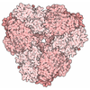


-Map
| File | Download / File: emd_56646.map.gz / Format: CCP4 / Size: 216 MB / Type: IMAGE STORED AS FLOATING POINT NUMBER (4 BYTES) | ||||||||||||||||||||||||||||||||||||
|---|---|---|---|---|---|---|---|---|---|---|---|---|---|---|---|---|---|---|---|---|---|---|---|---|---|---|---|---|---|---|---|---|---|---|---|---|---|
| Projections & slices | Image control
Images are generated by Spider. | ||||||||||||||||||||||||||||||||||||
| Voxel size | X=Y=Z: 0.85 Å | ||||||||||||||||||||||||||||||||||||
| Density |
| ||||||||||||||||||||||||||||||||||||
| Symmetry | Space group: 1 | ||||||||||||||||||||||||||||||||||||
| Details | EMDB XML:
|
 Z (Sec.)
 Y (Row.)
 X (Col.)


-Supplemental data
-Half map: Half map A
| File | emd_56646_half_map_1.map | ||||||||||||
|---|---|---|---|---|---|---|---|---|---|---|---|---|---|
| Annotation | Half map A | ||||||||||||
| Projections & Slices |
| ||||||||||||
| Density Histograms |


-Half map: Half map B
| File | emd_56646_half_map_2.map | ||||||||||||
|---|---|---|---|---|---|---|---|---|---|---|---|---|---|
| Annotation | Half map B | ||||||||||||
| Projections & Slices |
| ||||||||||||
| Density Histograms |


- Sample components
-Entire : UBA6-FAT10Dha-UBE2Z trapped ternary complex
| Entire | Name: UBA6-FAT10Dha-UBE2Z trapped ternary complex |
|---|---|
| Components |
|
-Supramolecule #1: UBA6-FAT10Dha-UBE2Z trapped ternary complex
| Supramolecule | Name: UBA6-FAT10Dha-UBE2Z trapped ternary complex / type: complex / ID: 1 / Parent: 0 / Macromolecule list: #1-#3 |
|---|---|
| Source (natural) | Organism: Homo sapiens (human) |
-Macromolecule #1: Ubiquitin-conjugating enzyme E2 Z
| Macromolecule | Name: Ubiquitin-conjugating enzyme E2 Z / type: protein_or_peptide / ID: 1 / Number of copies: 1 / Enantiomer: LEVO / EC number: E2 ubiquitin-conjugating enzyme |
|---|---|
| Source (natural) | Organism: Homo sapiens (human) |
| Molecular weight | Theoretical: 38.40325 KDa |
| Recombinant expression | Organism:   Escherichia coli (E. coli) Escherichia coli (E. coli) |
| Sequence | String: GPMAESPTEE AATAGAGAAG PGASSVAGVV GVSGSGGGFG PPFLPDVWAA AAAAGGAGGP GSGLAPLPGL PPSAAAHGAA LLSHWDPTL SSDWDGERTA PQCLLRIKRD IMSIYKEPPP GMFVVPDTVD MTKIHALITG PFDTPYEGGF FLFVFRCPPD Y PIHPPRVK ...String: GPMAESPTEE AATAGAGAAG PGASSVAGVV GVSGSGGGFG PPFLPDVWAA AAAAGGAGGP GSGLAPLPGL PPSAAAHGAA LLSHWDPTL SSDWDGERTA PQCLLRIKRD IMSIYKEPPP GMFVVPDTVD MTKIHALITG PFDTPYEGGF FLFVFRCPPD Y PIHPPRVK LMTTGNNTVR FNPNFYRNGK VCLSILGTWT GPAWSPAQSI SSVLISIQSL MTENPYHNEP GFEQERHPGD SK NYNECIR HETIRVAVCD MMEGKCPCPE PLRGVMEKSF LEYYDFYEVA CKDRLHLQGQ TMQDPFGEKR GHFDYQSLLM RLG LIRQKV LERLHNENAE MDSDSSSSGT ETDLHGSLRV UniProtKB: Ubiquitin-conjugating enzyme E2 Z |
-Macromolecule #2: Ubiquitin-like modifier-activating enzyme 6
| Macromolecule | Name: Ubiquitin-like modifier-activating enzyme 6 / type: protein_or_peptide / ID: 2 / Number of copies: 1 / Enantiomer: LEVO / EC number: E1 ubiquitin-activating enzyme |
|---|---|
| Source (natural) | Organism: Homo sapiens (human) |
| Molecular weight | Theoretical: 118.266883 KDa |
| Recombinant expression | Organism:   Spodoptera frugiperda (fall armyworm) Spodoptera frugiperda (fall armyworm) |
| Sequence | String: GPMEGSEPVA AHQGEEASCS SWGTGSTNKN LPIMSTASVE IDDALYSRQR YVLGDTAMQK MAKSHVFLSG MGGLGLEIAK NLVLAGIKA VTIHDTEKCQ AWDLGTNFFL SEDDVVNKRN RAEAVLKHIA ELNPYVHVTS SSVPFNETTD LSFLDKYQCV V LTEMKLPL ...String: GPMEGSEPVA AHQGEEASCS SWGTGSTNKN LPIMSTASVE IDDALYSRQR YVLGDTAMQK MAKSHVFLSG MGGLGLEIAK NLVLAGIKA VTIHDTEKCQ AWDLGTNFFL SEDDVVNKRN RAEAVLKHIA ELNPYVHVTS SSVPFNETTD LSFLDKYQCV V LTEMKLPL QKKINDFCRS QCPPIKFISA DVHGIWSRLF CDFGDEFEVL DTTGEEPKEI FISNITQANP GIVTCLENHP HK LETGQFL TFREINGMTG LNGSIQQITV ISPFSFSIGD TTELEPYLHG GIAVQVKTPK TVFFESLERQ LKHPKCLIVD FSN PEAPLE IHTAMLALDQ FQEKYSRKPN VGCQQDSEEL LKLATSISET LEEKPDVNAD IVHWLSWTAQ GFLSPLAAAV GGVA SQEVL KAVTGKFSPL CQWLYLEAAD IVESLGKPEC EEFLPRGDRY DALRACIGDT LCQKLQNLNI FLVGCGAIGC EMLKN FALL GVGTSKEKGM ITVTDPDLIE KSNLNRQFLF RPHHIQKPKS YTAADATLKI NSQIKIDAHL NKVCPTTETI YNDEFY TKQ DVIITALDNV EARRYVDSRC LANLRPLLDS GTMGTKGHTE VIVPHLTESY NSHRDPPEEE IPFCTLKSFP AAIEHTI QW ARDKFESSFS HKPSLFNKFW QTYSSAEEVL QKIQSGHSLE GCFQVIKLLS RRPRNWSQCV ELARLKFEKY FNHKALQL L HCFPLDIRLK DGSLFWQSPK RPPSPIKFDL NEPLHLSFLQ NAAKLYATVY CIPFAEEDLS ADALLNILSE VKIQEFKPS NKVVQTDETA RKPDHVPISS EDERNAIFQL EKAILSNEAT KSDLQMAVLS FEKDDDHNGH IDFITAASNL RAKMYSIEPA DRFKTKRIA GKIIPAIATT TATVSGLVAL EMIKVTGGYP FEAYKNCFLN LAIPIVVFTE TTEVRKTKIR NGISFTIWDR W TVHGKEDF TLLDFINAVK EKYGIEPTMV VQGVKMLYVP VMPGHAKRLK LTMHKLVKPT TEKKYVDLTV SFAPDIDGDE DL PGPPVRY YFSHDTD UniProtKB: Ubiquitin-like modifier-activating enzyme 6 |
-Macromolecule #3: Ubiquitin D
| Macromolecule | Name: Ubiquitin D / type: protein_or_peptide / ID: 3 / Number of copies: 1 / Enantiomer: LEVO |
|---|---|
| Source (natural) | Organism: Homo sapiens (human) |
| Molecular weight | Theoretical: 18.644664 KDa |
| Recombinant expression | Organism: Escherichia coli (E. coli) |
| Sequence | String: GPMAPNASTL TVHVRSEEWD LMTFDANPYD SVKKIKEHVR SKTKVPVQDQ VLLLGSKILK PRRSLSSYGI DKEKTIHLTL KVVKPSDEE LPLFLVESGD EAKRHLLQVR RSSSVAQVKA MIETKTGIIP ETQIVTLNGK RLEDGKMMAD YGIRKGNLLF L ASYSIGA UniProtKB: Ubiquitin D |
-Macromolecule #4: INOSITOL HEXAKISPHOSPHATE
| Macromolecule | Name: INOSITOL HEXAKISPHOSPHATE / type: ligand / ID: 4 / Number of copies: 1 / Formula: IHP |
|---|---|
| Molecular weight | Theoretical: 660.035 Da |
| Chemical component information | 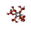 ChemComp-IHP: |
-Experimental details
-Structure determination
| Method | cryo EM |
|---|---|
 Processing Processing | single particle reconstruction |
| Aggregation state | particle |
-Sample preparation
| Buffer | pH: 7.5 |
|---|---|
| Vitrification | Cryogen name: ETHANE |
- Electron microscopy
| Microscope | TFS KRIOS |
|---|---|
| Image recording | Film or detector model: GATAN K3 BIOQUANTUM (6k x 4k) / Average electron dose: 42.7 e/Å2 |
| Electron beam | Acceleration voltage: 300 kV / Electron source:  FIELD EMISSION GUN FIELD EMISSION GUN |
| Electron optics | Illumination mode: FLOOD BEAM / Imaging mode: BRIGHT FIELD / Nominal defocus max: 2.5 µm / Nominal defocus min: 1.0 µm |
| Experimental equipment |  Model: Titan Krios / Image courtesy: FEI Company |

