 Movie
Movie Controller


+ Open data

- Basic information
| Entry | Database: PDB / ID: 28fu | |||||||||
|---|---|---|---|---|---|---|---|---|---|---|
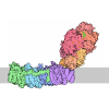
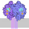
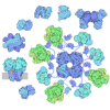
| Title | Cryo-EM structure of hydrogenase-2 in complex with L1 (right) | |||||||||
 Components Components |
| |||||||||
 Keywords Keywords | OXIDOREDUCTASE / Complex / Binder | |||||||||
| Function / homology |  Function and homology information Function and homology informationhydrogenase (acceptor) / ferredoxin hydrogenase complex / [Ni-Fe] hydrogenase complex / hydrogenase (acceptor) activity / ferredoxin hydrogenase activity / anaerobic respiration / nickel cation binding / iron-sulfur cluster binding / electron transfer activity / periplasmic space ...hydrogenase (acceptor) / ferredoxin hydrogenase complex / [Ni-Fe] hydrogenase complex / hydrogenase (acceptor) activity / ferredoxin hydrogenase activity / anaerobic respiration / nickel cation binding / iron-sulfur cluster binding / electron transfer activity / periplasmic space / membrane / plasma membrane Similarity search - Function | |||||||||
| Biological species | synthetic construct (others)  Escherichia coli (E. coli) Escherichia coli (E. coli) | |||||||||
| Method | ELECTRON MICROSCOPY / single particle reconstruction / cryo EM / Resolution: 2.48 Å | |||||||||
 Authors Authors | Zhang, N. / Huang, Y. / Zhang, L. | |||||||||
| Funding support |  China, 1items China, 1items
| |||||||||
 Citation Citation | Journal: Angew Chem Int Ed Engl / Year: 2026 Title: De Novo Design of a Protein Binder to Probe Gas Channel and Enhance the Oxygen Tolerance of [NiFe]-Hydrogenase. Authors: Xuan Sun / Qi Xiao / Nan Zhang / Wenjin Li / Hang Luo / Yanxin Gao / Leyan Zhang / Yilin Fan / Peiyu Jiang / Yucen Huang / Geng Wu / Liyun Zhang / Abstract: [NiFe]-Hydrogenases display remarkable catalytic efficiency for H production and oxidation but are sensitive to O, which severely restricts their biotechnological applications. The identities and ...[NiFe]-Hydrogenases display remarkable catalytic efficiency for H production and oxidation but are sensitive to O, which severely restricts their biotechnological applications. The identities and functional relevance of gas channels leading to the buried [NiFe] active site remain elusive, hindering rational enzyme engineering. Here, we introduce an artificial intelligence-guided de novo protein binder design strategy to map hydrophobic O diffusion pathways in Escherichia coli hydrogenase-2 (Hyd-2). By integrating RFdiffusion, ProteinMPNN, and AlphaFold, ∼100,000 candidate binders were computationally screened, yielding two high-affinity binders, L1 and L2. Biophysical and electrochemical analyses show that L1 selectively occludes the primary O ingress channel, enhancing the enzyme's oxygen tolerance by more than threefold, whereas L2, which targets another putative channel, has a negligible effect, indicating that this pathway contributes minimally to O ingress under the tested conditions. Furthermore, we determined the L1-Hyd-2 complex structure by cryo-electron microscopy and revealed the key interaction residues and interface conformation. Integrated structural and computational analyses provide mechanistic insights into the system's oxygen tolerance and high-affinity L1-Hyd-2 interaction. All findings provide the first direct experimental evidence for hierarchical O diffusion channels in [NiFe]-hydrogenases and establish binder-mediated channel occlusion as a generalizable, mutation-free strategy for elucidating and modulating gas transport in metalloenzymes. | |||||||||
| History |
|
- Structure visualization
| Structure viewer | Molecule: MolmilJmol/JSmol |
|---|
- Downloads & links
-Download
| PDBx/mmCIF format | 28fu.cif.gz | 342.1 KB | Display | PDBx/mmCIF format |
|---|---|---|---|---|
| PDB format | pdb28fu.ent.gz | 269 KB | Display | PDB format |
| PDBx/mmJSON format | 28fu.json.gz | Tree view | PDBx/mmJSON format | |
| Others |  Other downloads Other downloads |
-Validation report
| Arichive directory | https://data.pdbj.org/pub/pdb/validation_reports/8f/28fuftp://data.pdbj.org/pub/pdb/validation_reports/8f/28fu | HTTPS FTP |
|---|
-Related structure data
| Related structure data |  81713MC  28fsC M: map data used to model this data C: citing same article ( |
|---|---|
| Similar structure data |

-Links
 PDBj
PDBj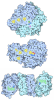

- Assembly
| Deposited unit | 
|
|---|---|
| 1 |
|
-Components
-Protein , 1 types, 1 molecules E
| #1: Protein | Mass: 8356.343 Da / Num. of mol.: 1 Source method: isolated from a genetically manipulated source Source: (gene. exp.) synthetic construct (others) / Production host: Escherichia coli (E. coli) |
|---|
-Hydrogenase-2 ... , 2 types, 4 molecules ACBD
| #2: Protein | Mass: 32371.498 Da / Num. of mol.: 2 Source method: isolated from a genetically manipulated source Source: (gene. exp.) Escherichia coli (strain K12) (bacteria)Strain: K12 / Gene: hybO, yghV, b2997, JW2965 / Production host: Escherichia coli (E. coli) / References: UniProt: P69741, hydrogenase (acceptor)#3: Protein | Mass: 62557.906 Da / Num. of mol.: 2 Source method: isolated from a genetically manipulated source Source: (gene. exp.) Escherichia coli (strain K12) (bacteria)Strain: K12 / Gene: hybC, b2994, JW2962 / Production host: Escherichia coli (E. coli) / References: UniProt: P0ACE0, hydrogenase (acceptor) |
|---|
-Non-polymers , 4 types, 10 molecules 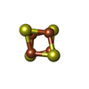
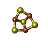
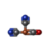

| #4: Chemical | ChemComp-SF4 / 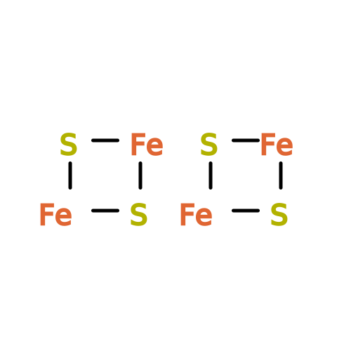 Mass: 351.640 Da / Num. of mol.: 4 / Source method: obtained synthetically / Formula: Fe4S4 Mass: 351.640 Da / Num. of mol.: 4 / Source method: obtained synthetically / Formula: Fe4S4#5: Chemical | 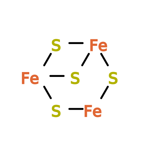 Mass: 295.795 Da / Num. of mol.: 2 / Source method: obtained synthetically / Formula: Fe3S4 Mass: 295.795 Da / Num. of mol.: 2 / Source method: obtained synthetically / Formula: Fe3S4#6: Chemical | 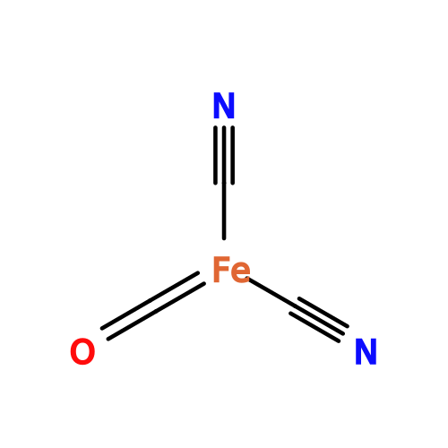 Mass: 135.890 Da / Num. of mol.: 2 / Source method: obtained synthetically / Formula: C3FeN2O Mass: 135.890 Da / Num. of mol.: 2 / Source method: obtained synthetically / Formula: C3FeN2O#7: Chemical |  Mass: 58.693 Da / Num. of mol.: 2 / Source method: obtained synthetically / Formula: Ni Mass: 58.693 Da / Num. of mol.: 2 / Source method: obtained synthetically / Formula: Ni |
|---|
-Details
| Has ligand of interest | N |
|---|---|
| Has protein modification | Y |
-Experimental details
-Experiment
| Experiment | Method: ELECTRON MICROSCOPY |
|---|---|
| EM experiment | Aggregation state: PARTICLE / 3D reconstruction method: single particle reconstruction |
- Sample preparation
| Component | Name: The complex of hydrogenase-2 with binder L1 (right) / Type: COMPLEX / Entity ID: #1-#3 / Source: RECOMBINANT |
|---|---|
| Source (natural) | Organism: Escherichia coli (strain K12) (bacteria) |
| Source (recombinant) | Organism: Escherichia coli (E. coli) |
| Buffer solution | pH: 7.2 |
| Specimen | Embedding applied: NO / Shadowing applied: NO / Staining applied: NO / Vitrification applied: YES |
| Vitrification | Cryogen name: ETHANE |
- Electron microscopy imaging
| Microscopy | Model: TFS TITAN THEMIS |
|---|---|
| Electron gun | Electron source:  FIELD EMISSION GUN / Accelerating voltage: 300 kV / Illumination mode: FLOOD BEAM FIELD EMISSION GUN / Accelerating voltage: 300 kV / Illumination mode: FLOOD BEAM |
| Electron lens | Mode: BRIGHT FIELD / Nominal defocus max: 2000 nm / Nominal defocus min: 800 nm |
| Image recording | Electron dose: 50 e/Å2 / Film or detector model: FEI FALCON IV (4k x 4k) |
- Processing
| EM software |
| ||||||||||||||||||||||||
|---|---|---|---|---|---|---|---|---|---|---|---|---|---|---|---|---|---|---|---|---|---|---|---|---|---|
| CTF correction | Type: NONE | ||||||||||||||||||||||||
| 3D reconstruction | Resolution: 2.48 Å / Resolution method: FSC 0.143 CUT-OFF / Num. of particles: 241696 / Symmetry type: POINT | ||||||||||||||||||||||||
| Refinement | Highest resolution: 2.48 Å Stereochemistry target values: REAL-SPACE (WEIGHTED MAP SUM AT ATOM CENTERS) | ||||||||||||||||||||||||
| Refine LS restraints |
|
