- PDB-26sj: Crystal structure of MAIT A-F7 TCR-MR1-xanthine -
+
Open data
ID or keywords:
Loading...
-
Basic information
Entry
Database: PDB / ID: 26sj
Title
Crystal structure of MAIT A-F7 TCR-MR1-xanthine
Components
(Human A-F7 TCR ...) x 2
Beta-2-microglobulin
Major histocompatibility complex class I-related gene protein
Keywords
IMMUNE SYSTEM / MAIT cells / MR1 / antigen presentation
Function / homology
Function and homology information
antigen processing and presentation of exogenous antigen / positive regulation of T cell mediated cytotoxicity directed against tumor cell target / MHC class I receptor activity / T cell differentiation in thymus / antigen processing and presentation of peptide antigen via MHC class I / beta-2-microglobulin binding / T cell receptor binding / regulation of natural killer cell mediated immunity / early endosome lumen / Nef mediated downregulation of MHC class I complex cell surface expression ...antigen processing and presentation of exogenous antigen / positive regulation of T cell mediated cytotoxicity directed against tumor cell target / MHC class I receptor activity / T cell differentiation in thymus / antigen processing and presentation of peptide antigen via MHC class I / beta-2-microglobulin binding / T cell receptor binding / regulation of natural killer cell mediated immunity / early endosome lumen / Nef mediated downregulation of MHC class I complex cell surface expression / DAP12 interactions / Endosomal/Vacuolar pathway / Antigen Presentation: Folding, assembly and peptide loading of class I MHC / regulation of iron ion transport / negative regulation of iron ion transport / negative regulation of forebrain neuron differentiation / antigen processing and presentation of exogenous peptide antigen via MHC class Ib / peptide antigen assembly with MHC class I protein complex / ER to Golgi transport vesicle membrane / HFE-transferrin receptor complex / MHC class I peptide loading complex / transferrin transport / negative regulation of receptor-mediated endocytosis / cellular response to iron ion / positive regulation of T cell cytokine production / antigen processing and presentation of endogenous peptide antigen via MHC class I / peptide antigen assembly with MHC class II protein complex / MHC class I protein complex / negative regulation of epithelial cell proliferation / cellular response to nicotine / negative regulation of neurogenesis / positive regulation of receptor-mediated endocytosis / MHC class II protein complex / specific granule lumen / positive regulation of immune response / antigen processing and presentation of exogenous peptide antigen via MHC class II / peptide antigen binding / recycling endosome membrane / phagocytic vesicle membrane / positive regulation of T cell activation / Interferon gamma signaling / Immunoregulatory interactions between a Lymphoid and a non-Lymphoid cell / sensory perception of smell / Modulation by Mtb of host immune system / tertiary granule lumen / positive regulation of cellular senescence / MHC class II protein complex binding / DAP12 signaling / late endosome membrane / ER-Phagosome pathway / early endosome membrane / defense response to Gram-negative bacterium / amyloid fibril formation / protein homotetramerization / intracellular iron ion homeostasis / learning or memory / defense response to Gram-positive bacterium / immune response / endoplasmic reticulum lumen / Amyloid fiber formation / external side of plasma membrane / Golgi membrane / innate immune response / focal adhesion / lysosomal membrane / Neutrophil degranulation / endoplasmic reticulum membrane / SARS-CoV-2 activates/modulates innate and adaptive immune responses / structural molecule activity / Golgi apparatus / endoplasmic reticulum / protein homodimerization activity / : / extracellular exosome / extracellular region / membrane / identical protein binding / plasma membrane Similarity search - Function
MHC class I alpha chain, alpha1 alpha2 domains / Class I Histocompatibility antigen, domains alpha 1 and 2 / Beta-2-Microglobulin / : / MHC class I-like antigen recognition-like / MHC class I-like antigen recognition-like superfamily / MHC classes I/II-like antigen recognition protein / : / Immunoglobulin/major histocompatibility complex, conserved site / Immunoglobulins and major histocompatibility complex proteins signature. ...MHC class I alpha chain, alpha1 alpha2 domains / Class I Histocompatibility antigen, domains alpha 1 and 2 / Beta-2-Microglobulin / : / MHC class I-like antigen recognition-like / MHC class I-like antigen recognition-like superfamily / MHC classes I/II-like antigen recognition protein / : / Immunoglobulin/major histocompatibility complex, conserved site / Immunoglobulins and major histocompatibility complex proteins signature. / Immunoglobulin C-Type / Immunoglobulin C1-set / Immunoglobulin C1-set domain / Ig-like domain profile. / Immunoglobulin-like domain / Immunoglobulin-like domain superfamily / Immunoglobulin-like fold Similarity search - Domain/homology
XANTHINE / Beta-2-microglobulin / Major histocompatibility complex class I-related protein 1 Similarity search - Component
A: Major histocompatibility complex class I-related gene protein B: Beta-2-microglobulin C: Major histocompatibility complex class I-related gene protein D: Human A-F7 TCR TRAV1-2_ALPHA E: Human A-F7 TCR TRBV6-1_BETA F: Beta-2-microglobulin G: Human A-F7 TCR TRAV1-2_ALPHA H: Human A-F7 TCR TRBV6-1_BETA hetero molecules
MajorhistocompatibilitycomplexclassI-relatedgeneprotein / MHC class I-related gene protein / Class I histocompatibility antigen-like protein
Mass: 31711.670 Da / Num. of mol.: 2 / Mutation: C261S Source method: isolated from a genetically manipulated source Source: (gene. exp.) Homo sapiens (human) / Gene: MR1 / Production host: Escherichia coli (E. coli) / References: UniProt: Q95460
#2: Protein
Beta-2-microglobulin
Mass: 11879.356 Da / Num. of mol.: 2 Source method: isolated from a genetically manipulated source Source: (gene. exp.) Homo sapiens (human) / Gene: B2M / Production host: Escherichia coli (E. coli) / References: UniProt: P61769
-
Human A-F7 TCR ... , 2 types, 4 molecules DGEH
#3: Protein
HumanA-F7TCRTRAV1-2_ALPHA
Mass: 22781.268 Da / Num. of mol.: 2 Source method: isolated from a genetically manipulated source Source: (gene. exp.) Homo sapiens (human) / Production host: Escherichia coli (E. coli)
#4: Protein
HumanA-F7TCRTRBV6-1_BETA
Mass: 27677.760 Da / Num. of mol.: 2 Source method: isolated from a genetically manipulated source Source: (gene. exp.) Homo sapiens (human) / Production host: Escherichia coli (E. coli)
In the structure databanks used in Yorodumi, some data are registered as the other names, "COVID-19 virus" and "2019-nCoV". Here are the details of the virus and the list of structure data.
Jan 31, 2019. EMDB accession codes are about to change! (news from PDBe EMDB page)
EMDB accession codes are about to change! (news from PDBe EMDB page)
The allocation of 4 digits for EMDB accession codes will soon come to an end. Whilst these codes will remain in use, new EMDB accession codes will include an additional digit and will expand incrementally as the available range of codes is exhausted. The current 4-digit format prefixed with “EMD-” (i.e. EMD-XXXX) will advance to a 5-digit format (i.e. EMD-XXXXX), and so on. It is currently estimated that the 4-digit codes will be depleted around Spring 2019, at which point the 5-digit format will come into force.
The EM Navigator/Yorodumi systems omit the EMD- prefix.
Related info.:Q: What is EMD? / ID/Accession-code notation in Yorodumi/EM Navigator
Yorodumi is a browser for structure data from EMDB, PDB, SASBDB, etc.
This page is also the successor to EM Navigator detail page, and also detail information page/front-end page for Omokage search.
The word "yorodu" (or yorozu) is an old Japanese word meaning "ten thousand". "mi" (miru) is to see.
Related info.:EMDB / PDB / SASBDB / Comparison of 3 databanks / Yorodumi Search / Aug 31, 2016. New EM Navigator & Yorodumi / Yorodumi Papers / Jmol/JSmol / Function and homology information / Changes in new EM Navigator and Yorodumi
 Movie
Movie Controller


 Open data
 Basic information
 Components
 Keywords
 Function and homology information
 Homo sapiens (human)
 X-RAY DIFFRACTION /
  Authors
 Australia, 1items
  Citation
 Structure visualization
 Downloads & links
 Other downloads

 PDBj
PDBj
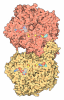
 Assembly

 Escherichia coli (E. coli) / References: UniProt: Q95460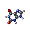
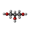



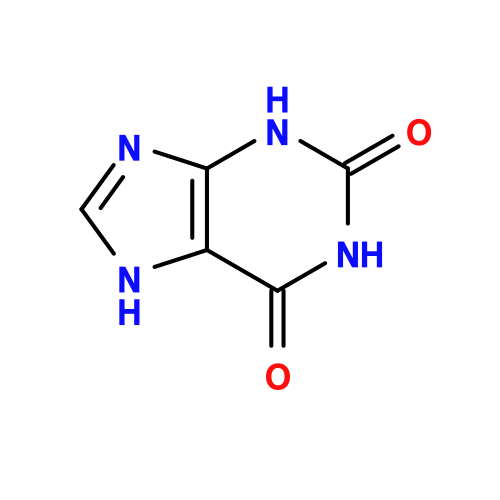 Mass: 152.111 Da / Num. of mol.: 2 / Source method: obtained synthetically / Formula: C5H4N4O2 / Feature type: SUBJECT OF INVESTIGATION
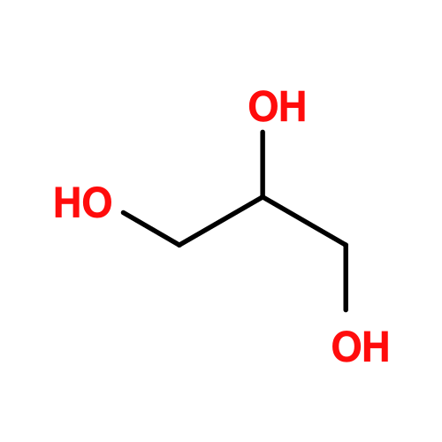 Mass: 92.094 Da / Num. of mol.: 8 / Source method: obtained synthetically / Formula: C3H8O3
 Mass: 35.453 Da / Num. of mol.: 2 / Source method: obtained synthetically / Formula: Cl
 Mass: 40.078 Da / Num. of mol.: 1 / Source method: obtained synthetically / Formula: Ca
 Sample preparation
 Processing