 Movie
Movie Controller

[English] 日本語
 Yorodumi
- PDB-252l: GENERATING LIGAND BINDING SITES IN T4 LYSOZYME USING DEFICIENCY-C... -
+ Open data

- Basic information
| Entry | Database: PDB / ID: 252l | ||||||
|---|---|---|---|---|---|---|---|
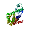
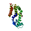
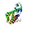
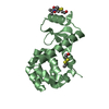
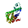
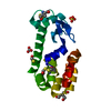
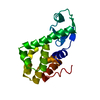
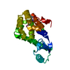
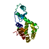
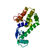
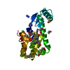
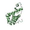
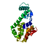
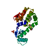
| Title | GENERATING LIGAND BINDING SITES IN T4 LYSOZYME USING DEFICIENCY-CREATING SUBSTITUTIONS | ||||||
 Components Components | T4 LYSOZYME | ||||||
 Keywords Keywords | HYDROLASE / O-GLYCOSYL / T4 LYSOZYME / CAVITY MUTANTS / LIGAND BINDING / PROTEIN ENGINEERING / PROTEIN DESIGN / MULTIPLE CONFORMATIONS | ||||||
| Function / homology |  Function and homology information Function and homology informationviral release from host cell by cytolysis / peptidoglycan catabolic process / cell wall macromolecule catabolic process / lysozyme / lysozyme activity / host cell cytoplasm / defense response to bacterium Similarity search - Function | ||||||
| Biological species |  Enterobacteria phage T4 (virus) Enterobacteria phage T4 (virus) | ||||||
| Method |  X-RAY DIFFRACTION / DIFFERENCE FOURIER / Resolution: 2.1 Å X-RAY DIFFRACTION / DIFFERENCE FOURIER / Resolution: 2.1 Å | ||||||
 Authors Authors | Baldwin, E.P. / Baase, W.A. / Zhang, X.-J. / Feher, V. / Matthews, B.W. | ||||||
 Citation Citation | Journal: J.Mol.Biol. / Year: 1998 Title: Generation of ligand binding sites in T4 lysozyme by deficiency-creating substitutions. Authors: Baldwin, E. / Baase, W.A. / Zhang, X. / Feher, V. / Matthews, B.W. #1: Journal: Nature / Year: 1992Title: A Cavity-Containing Mutant of T4 Lysozyme is Stabilized by Buried Benzene Authors: Eriksson, A.E. / Baase, W.A. / Wozniak, J.A. / Matthews, B.W. #2: Journal: Methods Enzymol. / Year: 1989Title: Expression and Nitrogen-15 Labeling of Proteins for Proton and Nitrogen-15 Nuclear Magnetic Resonance Authors: Muchmore, D.C. / Mcintosh, L.P. / Russell, C.B. / Anderson, D.E. / Dahlquist, F.W. #3: Journal: J.Mol.Biol. / Year: 1987Title: Structure of Bacteriophage T4 Lysozyme Refined at 1.7 A Resolution Authors: Weaver, L.H. / Matthews, B.W. | ||||||
| History |
|
- Structure visualization
| Structure viewer | Molecule: MolmilJmol/JSmol |
|---|
- Downloads & links
-Download
| PDBx/mmCIF format | 252l.cif.gz | 50.4 KB | Display | PDBx/mmCIF format |
|---|---|---|---|---|
| PDB format | pdb252l.ent.gz | 36.1 KB | Display | PDB format |
| PDBx/mmJSON format | 252l.json.gz | Tree view | PDBx/mmJSON format | |
| Others |  Other downloads Other downloads |
-Validation report
| Arichive directory | https://data.pdbj.org/pub/pdb/validation_reports/52/252lftp://data.pdbj.org/pub/pdb/validation_reports/52/252l | HTTPS FTP |
|---|
-Related structure data
| Related structure data | 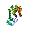 220lC 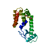 222lC 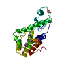 223lC  225lC 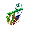 226lC 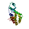 227lC  228lC 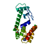 229lC C: citing same article ( |
|---|---|
| Similar structure data |


-Links
 PDBj
PDBj


- Assembly
| Deposited unit | 
| ||||||||
|---|---|---|---|---|---|---|---|---|---|
| 1 |
| ||||||||
| Unit cell |
|
-Components
| #1: Protein | Mass: 18508.125 Da / Num. of mol.: 1 / Mutation: C54T, C97A, M102A, M106A Source method: isolated from a genetically manipulated source Source: (gene. exp.) Enterobacteria phage T4 (virus) / Genus: T4-like viruses / Species: Enterobacteria phage T4 sensu lato / Cellular location: CYTOPLASM / Gene: GENE E / Plasmid: PHS1403 / Gene (production host): T4 LYSOZYME / Production host:  |
|---|---|
| #2: Chemical | ChemComp-CL /   Mass: 35.453 Da / Num. of mol.: 1 / Source method: obtained synthetically / Formula: Cl Mass: 35.453 Da / Num. of mol.: 1 / Source method: obtained synthetically / Formula: Cl |
| #3: Chemical | ChemComp-HED /   Mass: 154.251 Da / Num. of mol.: 1 / Source method: obtained synthetically / Formula: C4H10O2S2 Mass: 154.251 Da / Num. of mol.: 1 / Source method: obtained synthetically / Formula: C4H10O2S2 |
| #4: Water | ChemComp-HOH /  Mass: 18.015 Da / Num. of mol.: 136 / Source method: isolated from a natural source / Formula: H2O Mass: 18.015 Da / Num. of mol.: 136 / Source method: isolated from a natural source / Formula: H2O |
| Compound details | STRUCTURE OF A T4 LYSOZYME MUTANT THAT CONTAINS TWO DIFFERENT CONFORMATIONS FOR RESIDUES 106 - 114. ...STRUCTURE OF A T4 LYSOZYME MUTANT THAT CONTAINS TWO DIFFERENT CONFORMATI |
-Experimental details
-Experiment
| Experiment | Method: X-RAY DIFFRACTION / Number of used crystals: 1 |
|---|
- Sample preparation
| Crystal | Density Matthews: 2.8 Å3/Da / Density % sol: 56.1 % | ||||||||||||||||||||||||||||||||||||||||||||||||
|---|---|---|---|---|---|---|---|---|---|---|---|---|---|---|---|---|---|---|---|---|---|---|---|---|---|---|---|---|---|---|---|---|---|---|---|---|---|---|---|---|---|---|---|---|---|---|---|---|---|
| Crystal grow | Temperature: 277 K / Method: vapor diffusion, hanging drop / pH: 7 Details: CRYSTALS GROWN IN HANGING DROPS AT 4 DEGREES C. PROTEIN 10-20 MG/ML IN A BUFFER CONTAINING 0.5 M NACL 0.1 M NAPO4 PH 6.5 WAS DILUTED 1/2 WITH A WELL SOLUTION CONTAINING MIXED K/NAPO4 PH 6.3- ...Details: CRYSTALS GROWN IN HANGING DROPS AT 4 DEGREES C. PROTEIN 10-20 MG/ML IN A BUFFER CONTAINING 0.5 M NACL 0.1 M NAPO4 PH 6.5 WAS DILUTED 1/2 WITH A WELL SOLUTION CONTAINING MIXED K/NAPO4 PH 6.3-7.1,1.8-2.2 MOLAR., pH 7.0, vapor diffusion - hanging drop, temperature 277K PH range: 6.3-7.1 | ||||||||||||||||||||||||||||||||||||||||||||||||
| Crystal grow | *PLUS Method: batch method / Details: Remington, S.J., (1978). J. Mol. Biol., 118, 81. / pH: 6.7 | ||||||||||||||||||||||||||||||||||||||||||||||||
| Components of the solutions | *PLUS
|
-Data collection
| Diffraction | Mean temperature: 298 K |
|---|---|
| Diffraction source | Source: ROTATING ANODE / Type: RIGAKU RUH2R / Wavelength: 1.5418 |
| Detector | Type: XUONG-HAMLIN MULTIWIRE / Detector: AREA DETECTOR / Date: Mar 25, 1993 / Details: GRAPHITE MONOCHROMATOR |
| Radiation | Monochromator: GRAPHITE(002) / Monochromatic (M) / Laue (L): M / Scattering type: x-ray |
| Radiation wavelength | Wavelength: 1.5418 Å / Relative weight: 1 |
| Reflection | Resolution: 2.1→30 Å / Num. obs: 11176 / % possible obs: 85 % / Rmerge(I) obs: 0.049 |
| Reflection shell | Highest resolution: 2.1 Å / Rmerge(I) obs: 0.2 / Mean I/σ(I) obs: 2 |
- Processing
| Software |
| ||||||||||||||||||||||||||||||
|---|---|---|---|---|---|---|---|---|---|---|---|---|---|---|---|---|---|---|---|---|---|---|---|---|---|---|---|---|---|---|---|
| Refinement | Method to determine structure: DIFFERENCE FOURIER Starting model: CYS-FREE WILD TYPE LYSOZYME WITH RESIDUES 106 - 114 DELETED Resolution: 2.1→30 Å / σ(F): 0 / Stereochemistry target values: TNT PROTGEO Details: THE STARTING MODEL WAS CYS-FREE WILDTYPE WITH RESIDUES 106 - 115 DELETED AND 102 CONVERTED TO ALA. RESIDUES 106 - 114 WERE FIRST FIT IN A NON-WILDTYPE CONFORMATION. AFTER REFINEMENT THE ...Details: THE STARTING MODEL WAS CYS-FREE WILDTYPE WITH RESIDUES 106 - 115 DELETED AND 102 CONVERTED TO ALA. RESIDUES 106 - 114 WERE FIRST FIT IN A NON-WILDTYPE CONFORMATION. AFTER REFINEMENT THE WILDTYPE CONFORMATION WAS VISIBLE IN FO-FC DIFFERENCE MAPS, AND WAS ADDED TO THE MODEL. RELATIVE OCCUPANCIES WERE ESTIMATED BASED ON REFINED GROUP OCCUPANCIES (INITIALLY AT 0.5) AND EITHER FIXING OR REFINING THE B VALUES AND POSITIONS. THE WEIGHTS OF EACH CONFORMER ARE 0.6 (ALT) AND 0.4 (WT). IN MOST T4 MUTANT STRUCTURES, RESIDUES 163 AND 164 ARE DISORDERED AND NOT OBSERVED IN ELECTRON DENSITY MAPS. IN THIS MUTANT, THESE RESIDUES WERE RESOLVED IN REFINED FO-FC DIFFERENCE MAPS AND THE RESIDUES ADDED TO THE MODEL.
| ||||||||||||||||||||||||||||||
| Solvent computation | Solvent model: BABINET SCALING / Bsol: 630 Å2 / ksol: 1.1 e/Å3 | ||||||||||||||||||||||||||||||
| Refinement step | Cycle: LAST / Resolution: 2.1→30 Å
| ||||||||||||||||||||||||||||||
| Refine LS restraints |
| ||||||||||||||||||||||||||||||
| Software | *PLUS Name: TNT / Version: 5F / Classification: refinement | ||||||||||||||||||||||||||||||
| Refinement | *PLUS Rfactor all: 0.163 | ||||||||||||||||||||||||||||||
| Solvent computation | *PLUS | ||||||||||||||||||||||||||||||
| Displacement parameters | *PLUS | ||||||||||||||||||||||||||||||
| Refine LS restraints | *PLUS
|
