 Movie
Movie Controller


+ Open data

- Basic information
| Entry | Database: PDB / ID: 20zx | |||||||||||||||
|---|---|---|---|---|---|---|---|---|---|---|---|---|---|---|---|---|
| Title | Cryo-EM structure of Gq-coupled LPAR5 in complex with LPA | |||||||||||||||
 Components Components |
| |||||||||||||||
 Keywords Keywords | MEMBRANE PROTEIN / LPAR5 / LPA | |||||||||||||||
| Function / homology |  Function and homology information Function and homology informationLysosphingolipid and LPA receptors / G-protein activation / Activation of the phototransduction cascade / Glucagon-type ligand receptors / Thromboxane signalling through TP receptor / Sensory perception of sweet, bitter, and umami (glutamate) taste / G beta:gamma signalling through PI3Kgamma / G beta:gamma signalling through CDC42 / Cooperation of PDCL (PhLP1) and TRiC/CCT in G-protein beta folding / Activation of G protein gated Potassium channels ...Lysosphingolipid and LPA receptors / G-protein activation / Activation of the phototransduction cascade / Glucagon-type ligand receptors / Thromboxane signalling through TP receptor / Sensory perception of sweet, bitter, and umami (glutamate) taste / G beta:gamma signalling through PI3Kgamma / G beta:gamma signalling through CDC42 / Cooperation of PDCL (PhLP1) and TRiC/CCT in G-protein beta folding / Activation of G protein gated Potassium channels / Inhibition of voltage gated Ca2+ channels via Gbeta/gamma subunits / Ca2+ pathway / G alpha (z) signalling events / High laminar flow shear stress activates signaling by PIEZO1 and PECAM1:CDH5:KDR in endothelial cells / Glucagon-like Peptide-1 (GLP1) regulates insulin secretion / Vasopressin regulates renal water homeostasis via Aquaporins / Adrenaline,noradrenaline inhibits insulin secretion / ADP signalling through P2Y purinoceptor 12 / G alpha (q) signalling events / G alpha (i) signalling events / Thrombin signalling through proteinase activated receptors (PARs) / Activation of G protein gated Potassium channels / G-protein activation / G beta:gamma signalling through PI3Kgamma / Prostacyclin signalling through prostacyclin receptor / G beta:gamma signalling through PLC beta / ADP signalling through P2Y purinoceptor 1 / Thromboxane signalling through TP receptor / Presynaptic function of Kainate receptors / G beta:gamma signalling through CDC42 / Inhibition of voltage gated Ca2+ channels via Gbeta/gamma subunits / G alpha (12/13) signalling events / Glucagon-type ligand receptors / G beta:gamma signalling through BTK / ADP signalling through P2Y purinoceptor 12 / Adrenaline,noradrenaline inhibits insulin secretion / Cooperation of PDCL (PhLP1) and TRiC/CCT in G-protein beta folding / Ca2+ pathway / G alpha (z) signalling events / Thrombin signalling through proteinase activated receptors (PARs) / Extra-nuclear estrogen signaling / G alpha (q) signalling events / G alpha (s) signalling events / photoreceptor outer segment membrane / spectrin binding / Glucagon-like Peptide-1 (GLP1) regulates insulin secretion / G alpha (i) signalling events / High laminar flow shear stress activates signaling by PIEZO1 and PECAM1:CDH5:KDR in endothelial cells / Vasopressin regulates renal water homeostasis via Aquaporins / sensory perception of taste / alkylglycerophosphoethanolamine phosphodiesterase activity / retina development in camera-type eye / behavioral response to pain / photoreceptor outer segment / cardiac muscle cell apoptotic process / photoreceptor inner segment / cell population proliferation / G protein-coupled receptor activity / phospholipase C-activating G protein-coupled receptor signaling pathway / cellular response to catecholamine stimulus / adenylate cyclase-activating dopamine receptor signaling pathway / cellular response to prostaglandin E stimulus / heterotrimeric G-protein complex / G-protein beta-subunit binding / positive regulation of cytosolic calcium ion concentration / signaling receptor complex adaptor activity / cell body / GTPase binding / cellular response to hypoxia / G alpha (i) signalling events / G alpha (q) signalling events / G protein-coupled receptor signaling pathway / GTPase activity / synapse / dendrite / protein-containing complex binding / membrane / plasma membrane / cytoplasm Similarity search - Function | |||||||||||||||
| Biological species |   Rattus norvegicus (Norway rat) Rattus norvegicus (Norway rat) Bos taurus (domestic cattle) Bos taurus (domestic cattle) Homo sapiens (human) Homo sapiens (human) | |||||||||||||||
| Method | ELECTRON MICROSCOPY / single particle reconstruction / cryo EM / Resolution: 2.96 Å | |||||||||||||||
 Authors Authors | Zhao, L. / Li, X. | |||||||||||||||
| Funding support |  China, 2items China, 2items
| |||||||||||||||
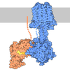
 Citation Citation | Journal: Proc Natl Acad Sci U S A / Year: 2026 Title: Structural basis for lysophosphatidic acid recognition and atypical Gα coupling by LPAR5. Authors: Xin Li / Kai Wang / Zhongliang Xing / Min Zhang / Wen Hu / Qingning Yuan / H Eric Xu / Li-Hua Zhao / Abstract: Lysophosphatidic acid receptor 5 (LPAR5) is a non-endothelial differentiation gene class A G protein-coupled receptor that regulates neuropathic pain, itch, and cancer progression through coupling to ...Lysophosphatidic acid receptor 5 (LPAR5) is a non-endothelial differentiation gene class A G protein-coupled receptor that regulates neuropathic pain, itch, and cancer progression through coupling to G proteins. Here, we report the cryo-EM structure of LPAR5 bound to 1-oleoyl-lysophosphatidic acid (LPA) in complex with G at 2.96 Å resolution, revealing a distinct mode of receptor activation and G protein coupling. The phosphate headgroup of LPA forms extensive polar interactions with residues from extracellular loop 2 and transmembrane helices TM5-TM7, while the lipid tail inserts into a deep hydrophobic cavity formed by TM3-TM5. Site-directed mutagenesis confirms the functional importance of these interactions. Remarkably, LPAR5 exhibits a noncanonical G protein coupling mode. Unlike previously reported GPCR-G protein structures in which the Gα C-terminal α5 helix ("wavy hook") primarily engages TM6, the wavy hook in LPAR5 is positioned toward the intracellular loop 1-helix 8 interface. This configuration is associated with limited TM6 outward displacement and modest rearrangement at the toggle-switch position (6.48). The resulting interface is stabilized by receptor-specific interactions and supported by functional data. Together, these findings reveal an alternative mode of GPCR-G protein coupling and highlight the structural plasticity underlying signaling specificity in LPA receptors. | |||||||||||||||
| History |
|
- Structure visualization
| Structure viewer | Molecule: MolmilJmol/JSmol |
|---|
- Downloads & links
-Download
| PDBx/mmCIF format | 20zx.cif.gz | 241.9 KB | Display | PDBx/mmCIF format |
|---|---|---|---|---|
| PDB format | pdb20zx.ent.gz | 187.5 KB | Display | PDB format |
| PDBx/mmJSON format | 20zx.json.gz | Tree view | PDBx/mmJSON format | |
| Others |  Other downloads Other downloads |
-Validation report
| Arichive directory | https://data.pdbj.org/pub/pdb/validation_reports/0z/20zxftp://data.pdbj.org/pub/pdb/validation_reports/0z/20zx | HTTPS FTP |
|---|
-Related structure data
| Related structure data |  67439MC M: map data used to model this data C: citing same article ( |
|---|---|
| Similar structure data |


-Links
 PDBj
PDBj











- Assembly
| Deposited unit | 
|
|---|---|
| 1 |
|
-Components
-Guanine nucleotide-binding protein ... , 3 types, 3 molecules BGA
| #1: Protein | Mass: 40226.992 Da / Num. of mol.: 1 Source method: isolated from a genetically manipulated source Source: (gene. exp.) Rattus norvegicus (Norway rat) / Gene: Gnb1 / Production host:  Trichoplusia ni (cabbage looper) / References: UniProt: P54311 Trichoplusia ni (cabbage looper) / References: UniProt: P54311 |
|---|---|
| #2: Protein | Mass: 7729.947 Da / Num. of mol.: 1 Source method: isolated from a genetically manipulated source Source: (gene. exp.) Bos taurus (domestic cattle) / Gene: GNG2 / Production host: Trichoplusia ni (cabbage looper) / References: UniProt: P63212 |
| #4: Protein | Mass: 41708.383 Da / Num. of mol.: 1 Source method: isolated from a genetically manipulated source Source: (gene. exp.) Homo sapiens (human) / Production host: Trichoplusia ni (cabbage looper) |
-Protein / Antibody / Non-polymers , 3 types, 3 molecules RE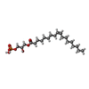
| #3: Protein | Mass: 41396.574 Da / Num. of mol.: 1 Source method: isolated from a genetically manipulated source Source: (gene. exp.) Homo sapiens (human) / Gene: LPAR5, GPR92, GPR93 / Production host: Trichoplusia ni (cabbage looper) / References: UniProt: Q9H1C0 |
|---|---|
| #5: Antibody | Mass: 26277.299 Da / Num. of mol.: 1 Source method: isolated from a genetically manipulated source Source: (gene. exp.) Homo sapiens (human) / Production host: Trichoplusia ni (cabbage looper) |
| #6: Chemical | ChemComp-UBL / [(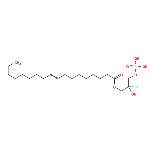 Mass: 436.520 Da / Num. of mol.: 1 / Source method: obtained synthetically / Formula: C21H41O7P / Feature type: SUBJECT OF INVESTIGATION Mass: 436.520 Da / Num. of mol.: 1 / Source method: obtained synthetically / Formula: C21H41O7P / Feature type: SUBJECT OF INVESTIGATION |
-Details
| Has ligand of interest | Y |
|---|---|
| Has protein modification | Y |
-Experimental details
-Experiment
| Experiment | Method: ELECTRON MICROSCOPY |
|---|---|
| EM experiment | Aggregation state: PARTICLE / 3D reconstruction method: single particle reconstruction |
- Sample preparation
| Component | Name: Cryo-EM structure of Gq-coupled LPAR5 in complex with LPA Type: COMPLEX / Entity ID: #1-#5 / Source: RECOMBINANT |
|---|---|
| Source (natural) | Organism: Homo sapiens (human) |
| Source (recombinant) | Organism: Trichoplusia ni (cabbage looper) |
| Buffer solution | pH: 7.4 |
| Specimen | Embedding applied: NO / Shadowing applied: NO / Staining applied: NO / Vitrification applied: YES |
| Vitrification | Cryogen name: ETHANE |
- Electron microscopy imaging
| Experimental equipment |  Model: Titan Krios / Image courtesy: FEI Company |
|---|---|
| Microscopy | Model: TFS KRIOS |
| Electron gun | Electron source:  FIELD EMISSION GUN / Accelerating voltage: 300 kV / Illumination mode: FLOOD BEAM FIELD EMISSION GUN / Accelerating voltage: 300 kV / Illumination mode: FLOOD BEAM |
| Electron lens | Mode: BRIGHT FIELD / Nominal defocus max: 18000 nm / Nominal defocus min: 8000 nm |
| Image recording | Electron dose: 50 e/Å2 / Film or detector model: FEI FALCON IV (4k x 4k) |
- Processing
| EM software |
| ||||||||||||||||||||||||
|---|---|---|---|---|---|---|---|---|---|---|---|---|---|---|---|---|---|---|---|---|---|---|---|---|---|
| CTF correction | Type: PHASE FLIPPING AND AMPLITUDE CORRECTION | ||||||||||||||||||||||||
| 3D reconstruction | Resolution: 2.96 Å / Resolution method: FSC 0.143 CUT-OFF / Num. of particles: 102163 / Symmetry type: POINT | ||||||||||||||||||||||||
| Refinement | Highest resolution: 2.96 Å Stereochemistry target values: REAL-SPACE (WEIGHTED MAP SUM AT ATOM CENTERS) | ||||||||||||||||||||||||
| Refine LS restraints |
|
