 Movie
Movie Controller


+ Open data

- Basic information
| Entry |  | |||||||||
|---|---|---|---|---|---|---|---|---|---|---|
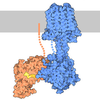
| Title | Cryo-EM structure of Gq-coupled LPAR5 in complex with LPA | |||||||||
 Map data Map data | ||||||||||
 Sample Sample |
| |||||||||
 Keywords Keywords | LPAR5 / LPA / MEMBRANE PROTEIN | |||||||||
| Function / homology |  Function and homology information Function and homology informationLysosphingolipid and LPA receptors / G-protein activation / Activation of the phototransduction cascade / Glucagon-type ligand receptors / Thromboxane signalling through TP receptor / Sensory perception of sweet, bitter, and umami (glutamate) taste / G beta:gamma signalling through PI3Kgamma / G beta:gamma signalling through CDC42 / Cooperation of PDCL (PhLP1) and TRiC/CCT in G-protein beta folding / Activation of G protein gated Potassium channels ...Lysosphingolipid and LPA receptors / G-protein activation / Activation of the phototransduction cascade / Glucagon-type ligand receptors / Thromboxane signalling through TP receptor / Sensory perception of sweet, bitter, and umami (glutamate) taste / G beta:gamma signalling through PI3Kgamma / G beta:gamma signalling through CDC42 / Cooperation of PDCL (PhLP1) and TRiC/CCT in G-protein beta folding / Activation of G protein gated Potassium channels / Inhibition of voltage gated Ca2+ channels via Gbeta/gamma subunits / Ca2+ pathway / G alpha (z) signalling events / High laminar flow shear stress activates signaling by PIEZO1 and PECAM1:CDH5:KDR in endothelial cells / Glucagon-like Peptide-1 (GLP1) regulates insulin secretion / Vasopressin regulates renal water homeostasis via Aquaporins / Adrenaline,noradrenaline inhibits insulin secretion / ADP signalling through P2Y purinoceptor 12 / G alpha (q) signalling events / G alpha (i) signalling events / Thrombin signalling through proteinase activated receptors (PARs) / Activation of G protein gated Potassium channels / G-protein activation / G beta:gamma signalling through PI3Kgamma / Prostacyclin signalling through prostacyclin receptor / G beta:gamma signalling through PLC beta / ADP signalling through P2Y purinoceptor 1 / Thromboxane signalling through TP receptor / Presynaptic function of Kainate receptors / G beta:gamma signalling through CDC42 / Inhibition of voltage gated Ca2+ channels via Gbeta/gamma subunits / G alpha (12/13) signalling events / Glucagon-type ligand receptors / G beta:gamma signalling through BTK / ADP signalling through P2Y purinoceptor 12 / Adrenaline,noradrenaline inhibits insulin secretion / Cooperation of PDCL (PhLP1) and TRiC/CCT in G-protein beta folding / Ca2+ pathway / G alpha (z) signalling events / Thrombin signalling through proteinase activated receptors (PARs) / Extra-nuclear estrogen signaling / G alpha (q) signalling events / G alpha (s) signalling events / photoreceptor outer segment membrane / spectrin binding / Glucagon-like Peptide-1 (GLP1) regulates insulin secretion / G alpha (i) signalling events / High laminar flow shear stress activates signaling by PIEZO1 and PECAM1:CDH5:KDR in endothelial cells / Vasopressin regulates renal water homeostasis via Aquaporins / sensory perception of taste / alkylglycerophosphoethanolamine phosphodiesterase activity / retina development in camera-type eye / behavioral response to pain / photoreceptor outer segment / cardiac muscle cell apoptotic process / photoreceptor inner segment / cell population proliferation / G protein-coupled receptor activity / phospholipase C-activating G protein-coupled receptor signaling pathway / cellular response to catecholamine stimulus / adenylate cyclase-activating dopamine receptor signaling pathway / cellular response to prostaglandin E stimulus / heterotrimeric G-protein complex / G-protein beta-subunit binding / positive regulation of cytosolic calcium ion concentration / signaling receptor complex adaptor activity / cell body / GTPase binding / cellular response to hypoxia / G alpha (i) signalling events / G alpha (q) signalling events / G protein-coupled receptor signaling pathway / GTPase activity / synapse / dendrite / protein-containing complex binding / membrane / plasma membrane / cytoplasm Similarity search - Function | |||||||||
| Biological species |  Homo sapiens (human) / Homo sapiens (human) /   Rattus norvegicus (Norway rat) / Rattus norvegicus (Norway rat) /  Bos taurus (domestic cattle) Bos taurus (domestic cattle) | |||||||||
| Method | single particle reconstruction / cryo EM / Resolution: 2.96 Å | |||||||||
 Authors Authors | Zhao L / Li X | |||||||||
| Funding support |  China, 2 items China, 2 items
| |||||||||
 Citation Citation | Journal: Proc Natl Acad Sci U S A / Year: 2026 Title: Structural basis for lysophosphatidic acid recognition and atypical Gα coupling by LPAR5. Authors: Xin Li / Kai Wang / Zhongliang Xing / Min Zhang / Wen Hu / Qingning Yuan / H Eric Xu / Li-Hua Zhao / Abstract: Lysophosphatidic acid receptor 5 (LPAR5) is a non-endothelial differentiation gene class A G protein-coupled receptor that regulates neuropathic pain, itch, and cancer progression through coupling to ...Lysophosphatidic acid receptor 5 (LPAR5) is a non-endothelial differentiation gene class A G protein-coupled receptor that regulates neuropathic pain, itch, and cancer progression through coupling to G proteins. Here, we report the cryo-EM structure of LPAR5 bound to 1-oleoyl-lysophosphatidic acid (LPA) in complex with G at 2.96 Å resolution, revealing a distinct mode of receptor activation and G protein coupling. The phosphate headgroup of LPA forms extensive polar interactions with residues from extracellular loop 2 and transmembrane helices TM5-TM7, while the lipid tail inserts into a deep hydrophobic cavity formed by TM3-TM5. Site-directed mutagenesis confirms the functional importance of these interactions. Remarkably, LPAR5 exhibits a noncanonical G protein coupling mode. Unlike previously reported GPCR-G protein structures in which the Gα C-terminal α5 helix ("wavy hook") primarily engages TM6, the wavy hook in LPAR5 is positioned toward the intracellular loop 1-helix 8 interface. This configuration is associated with limited TM6 outward displacement and modest rearrangement at the toggle-switch position (6.48). The resulting interface is stabilized by receptor-specific interactions and supported by functional data. Together, these findings reveal an alternative mode of GPCR-G protein coupling and highlight the structural plasticity underlying signaling specificity in LPA receptors. | |||||||||
| History |
|
- Structure visualization
| Supplemental images |
|---|

- Downloads & links
-EMDB archive
| Map data | emd_67439.map.gz | 166.3 MB | EMDB map data format | |
|---|---|---|---|---|
| Header (meta data) | emd-67439-v30.xmlemd-67439.xml | 22 KB 22 KB | Display Display | EMDB header |
| Images |  emd_67439.png emd_67439.png | 71.2 KB | ||
| Filedesc metadata | emd-67439.cif.gz | 7 KB | ||
| Archive directory |  http://ftp.pdbj.org/pub/emdb/structures/EMD-67439ftp://ftp.pdbj.org/pub/emdb/structures/EMD-67439 http://ftp.pdbj.org/pub/emdb/structures/EMD-67439ftp://ftp.pdbj.org/pub/emdb/structures/EMD-67439 | HTTPS FTP |
-Related structure data
| Related structure data |  20zxMC M: atomic model generated by this map C: citing same article ( |
|---|---|
| Similar structure data |


-Links
| EMDB pages | EMDB (EBI/PDBe) / EMDataResource |
|---|---|
| Related items in Molecule of the Month |










-Map
| File | Download / File: emd_67439.map.gz / Format: CCP4 / Size: 178 MB / Type: IMAGE STORED AS FLOATING POINT NUMBER (4 BYTES) | ||||||||||||||||||||||||||||||||||||
|---|---|---|---|---|---|---|---|---|---|---|---|---|---|---|---|---|---|---|---|---|---|---|---|---|---|---|---|---|---|---|---|---|---|---|---|---|---|
| Projections & slices | Image control
Images are generated by Spider. | ||||||||||||||||||||||||||||||||||||
| Voxel size | X=Y=Z: 0.73 Å | ||||||||||||||||||||||||||||||||||||
| Density |
| ||||||||||||||||||||||||||||||||||||
| Symmetry | Space group: 1 | ||||||||||||||||||||||||||||||||||||
| Details | EMDB XML:
|
 Z (Sec.)
 Y (Row.)
 X (Col.)


-Supplemental data
- Sample components
-Entire : Cryo-EM structure of Gq-coupled LPAR5 in complex with LPA
| Entire | Name: Cryo-EM structure of Gq-coupled LPAR5 in complex with LPA |
|---|---|
| Components |
|
-Supramolecule #1: Cryo-EM structure of Gq-coupled LPAR5 in complex with LPA
| Supramolecule | Name: Cryo-EM structure of Gq-coupled LPAR5 in complex with LPA type: complex / ID: 1 / Parent: 0 / Macromolecule list: #1-#5 |
|---|---|
| Source (natural) | Organism: Homo sapiens (human) |
-Macromolecule #1: Guanine nucleotide-binding protein G(I)/G(S)/G(T) subunit beta-1
| Macromolecule | Name: Guanine nucleotide-binding protein G(I)/G(S)/G(T) subunit beta-1 type: protein_or_peptide / ID: 1 / Number of copies: 1 / Enantiomer: LEVO |
|---|---|
| Source (natural) | Organism: Rattus norvegicus (Norway rat) |
| Molecular weight | Theoretical: 40.226992 KDa |
| Recombinant expression | Organism:  Trichoplusia ni (cabbage looper) Trichoplusia ni (cabbage looper) |
| Sequence | String: MGSLLQSELD QLRQEAEQLK NQIRDARKAC ADATLSQITN NIDPVGRIQM RTRRTLRGHL AKIYAMHWGT DSRLLVSASQ DGKLIIWDS YTTNKVHAIP LRSSWVMTCA YAPSGNYVAC GGLDNICSIY NLKTREGNVR VSRELAGHTG YLSCCRFLDD N QIVTSSGD ...String: MGSLLQSELD QLRQEAEQLK NQIRDARKAC ADATLSQITN NIDPVGRIQM RTRRTLRGHL AKIYAMHWGT DSRLLVSASQ DGKLIIWDS YTTNKVHAIP LRSSWVMTCA YAPSGNYVAC GGLDNICSIY NLKTREGNVR VSRELAGHTG YLSCCRFLDD N QIVTSSGD TTCALWDIET GQQTTTFTGH TGDVMSLSLA PDTRLFVSGA CDASAKLWDV REGMCRQTFT GHESDINAIC FF PNGNAFA TGSDDATCRL FDLRADQELM TYSHDNIICG ITSVSFSKSG RLLLAGYDDF NCNVWDALKA DRAGVLAGHD NRV SCLGVT DDGMAVATGS WDSFLKIWNG SSGGGGSGGG GSSGVSGWRL FKKIS UniProtKB: Guanine nucleotide-binding protein G(I)/G(S)/G(T) subunit beta-1 |
-Macromolecule #2: Guanine nucleotide-binding protein G(I)/G(S)/G(O) subunit gamma-2
| Macromolecule | Name: Guanine nucleotide-binding protein G(I)/G(S)/G(O) subunit gamma-2 type: protein_or_peptide / ID: 2 / Number of copies: 1 / Enantiomer: LEVO |
|---|---|
| Source (natural) | Organism: Bos taurus (domestic cattle) |
| Molecular weight | Theoretical: 7.729947 KDa |
| Recombinant expression | Organism: Trichoplusia ni (cabbage looper) |
| Sequence | String: ASNNTASIAQ ARKLVEQLKM EANIDRIKVS KAAADLMAYC EAHAKEDPLL TPVPASENPF REKKFFCAIL UniProtKB: Guanine nucleotide-binding protein G(I)/G(S)/G(O) subunit gamma-2 |
-Macromolecule #3: Lysophosphatidic acid receptor 5
| Macromolecule | Name: Lysophosphatidic acid receptor 5 / type: protein_or_peptide / ID: 3 / Number of copies: 1 / Enantiomer: LEVO |
|---|---|
| Source (natural) | Organism: Homo sapiens (human) |
| Molecular weight | Theoretical: 41.396574 KDa |
| Recombinant expression | Organism: Trichoplusia ni (cabbage looper) |
| Sequence | String: MLANSSSTNS SVLPCPDYRP THRLHLVVYS LVLAAGLPLN ALALWVFLRA LRVHSVVSVY MCNLAASDLL FTLSLPVRLS YYALHHWPF PDLLCQTTGA IFQMNMYGSC IFLMLINVDR YAAIVHPLRL RHLRRPRVAR LLCLGVWALI LVFAVPAARV H RPSRCRYR ...String: MLANSSSTNS SVLPCPDYRP THRLHLVVYS LVLAAGLPLN ALALWVFLRA LRVHSVVSVY MCNLAASDLL FTLSLPVRLS YYALHHWPF PDLLCQTTGA IFQMNMYGSC IFLMLINVDR YAAIVHPLRL RHLRRPRVAR LLCLGVWALI LVFAVPAARV H RPSRCRYR DLEVRLCFES FSDELWKGRL LPLVLLAEAL GFLLPLAAVV YSSGRVFWTL ARPDATQSQR RRKTVRLLLA NL VIFLLCF VPYNSTLAVY GLLRSKLVAA SVPARDRVRG VLMVMVLLAG ANCVLDPLVY YFSAEGFRNT LRGLGTPHRA RTS ATNGTR AALAQSERSA VTTDATRPDA ASQGLLRPSD SHSLSSFTQC PQDSAL UniProtKB: Lysophosphatidic acid receptor 5 |
-Macromolecule #4: Guanine nucleotide-binding protein G(324) subunit alpha
| Macromolecule | Name: Guanine nucleotide-binding protein G(324) subunit alpha type: protein_or_peptide / ID: 4 / Number of copies: 1 / Enantiomer: LEVO |
|---|---|
| Source (natural) | Organism: Homo sapiens (human) |
| Molecular weight | Theoretical: 41.708383 KDa |
| Recombinant expression | Organism: Trichoplusia ni (cabbage looper) |
| Sequence | String: MGCTLSAEDK AAVERSKMIE KQLQKDKQVY RRTLRLLLLG ADNSGKSTIV KQMRIYHVNG YSEEECKQYK AVVYSNTIQS IIAIIRAMG RLKIDFGDSA RADDARQLFV LAGAAEEGFM TAELAGVIKR LWKDSGVQAC FNRSREYQLN DSAAYYLNDL D RIAQPNYI ...String: MGCTLSAEDK AAVERSKMIE KQLQKDKQVY RRTLRLLLLG ADNSGKSTIV KQMRIYHVNG YSEEECKQYK AVVYSNTIQS IIAIIRAMG RLKIDFGDSA RADDARQLFV LAGAAEEGFM TAELAGVIKR LWKDSGVQAC FNRSREYQLN DSAAYYLNDL D RIAQPNYI PTQQDVLRTR VKTSGIFETK FQVDKVNFHM FDVGAQRDER RKWIQCFNDV TAIIFVVDSS DYNRLQEALN DF KSIWNNR WLRTISVILF LNKQDLLAEK VLAGKSKIED YFPEFARYTT PEDATPEPGE DPRVTRAKYF IRKEFVDIST ASG DGRHIC YPHFTCAVDT ENARRIFNDC KDIILQMNLR EYNLV |
-Macromolecule #5: scFv16
| Macromolecule | Name: scFv16 / type: protein_or_peptide / ID: 5 / Number of copies: 1 / Enantiomer: LEVO |
|---|---|
| Source (natural) | Organism: Homo sapiens (human) |
| Molecular weight | Theoretical: 26.277299 KDa |
| Recombinant expression | Organism: Trichoplusia ni (cabbage looper) |
| Sequence | String: VQLVESGGGL VQPGGSRKLS CSASGFAFSS FGMHWVRQAP EKGLEWVAYI SSGSGTIYYA DTVKGRFTIS RDDPKNTLFL QMTSLRSED TAMYYCVRSI YYYGSSPFDF WGQGTTLTVS AGGGGSGGGG SGGGGSADIV MTQATSSVPV TPGESVSISC R SSKSLLHS ...String: VQLVESGGGL VQPGGSRKLS CSASGFAFSS FGMHWVRQAP EKGLEWVAYI SSGSGTIYYA DTVKGRFTIS RDDPKNTLFL QMTSLRSED TAMYYCVRSI YYYGSSPFDF WGQGTTLTVS AGGGGSGGGG SGGGGSADIV MTQATSSVPV TPGESVSISC R SSKSLLHS NGNTYLYWFL QRPGQSPQLL IYRMSNLASG VPDRFSGSGS GTAFTLTISR LEAEDVGVYY CMQHLEYPLT FG AGTKLEL |
-Macromolecule #6: [(2R)-2-oxidanyl-3-phosphonooxy-propyl] (Z)-octadec-9-enoate
| Macromolecule | Name: [(2R)-2-oxidanyl-3-phosphonooxy-propyl] (Z)-octadec-9-enoate type: ligand / ID: 6 / Number of copies: 1 / Formula: UBL |
|---|---|
| Molecular weight | Theoretical: 436.52 Da |
| Chemical component information | 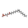 ChemComp-UBL: |
-Experimental details
-Structure determination
| Method | cryo EM |
|---|---|
 Processing Processing | single particle reconstruction |
| Aggregation state | particle |
-Sample preparation
| Buffer | pH: 7.4 |
|---|---|
| Vitrification | Cryogen name: ETHANE |
- Electron microscopy
| Microscope | TFS KRIOS |
|---|---|
| Image recording | Film or detector model: FEI FALCON IV (4k x 4k) / Average electron dose: 50.0 e/Å2 |
| Electron beam | Acceleration voltage: 300 kV / Electron source:  FIELD EMISSION GUN FIELD EMISSION GUN |
| Electron optics | Illumination mode: FLOOD BEAM / Imaging mode: BRIGHT FIELD / Nominal defocus max: 18.0 µm / Nominal defocus min: 8.0 µm |
| Experimental equipment |  Model: Titan Krios / Image courtesy: FEI Company |
