[English] 日本語
 Yorodumi
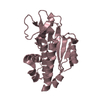
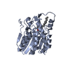
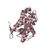
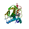
- PDB-1zrr: Residual Dipolar Coupling Refinement of Acireductone Dioxygenase ... -
+ Open data
- Basic information
| Entry | Database: PDB / ID: 1zrr | |||||||||
|---|---|---|---|---|---|---|---|---|---|---|
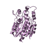
| Title | Residual Dipolar Coupling Refinement of Acireductone Dioxygenase from Klebsiella | |||||||||
 Components Components | E-2/E-2' protein | |||||||||
 Keywords Keywords | OXIDOREDUCTASE / nickel / cupin / beta helix / methionine salvage | |||||||||
| Function / homology |  Function and homology information Function and homology informationacireductone dioxygenase (Ni2+-requiring) / acireductone dioxygenase [iron(II)-requiring] / acireductone dioxygenase (Ni2+-requiring) activity / acireductone dioxygenase [iron(II)-requiring] activity / L-methionine salvage from S-adenosylmethionine / L-methionine salvage from methylthioadenosine / nickel cation binding / iron ion binding Similarity search - Function | |||||||||
| Biological species |  Klebsiella oxytoca (bacteria) Klebsiella oxytoca (bacteria) | |||||||||
| Method | SOLUTION NMR / combined torsional, cartesian dynamics simulated annealing with residual dipolar couplings, NOE, chemical shift, dihedral restraints | |||||||||
 Authors Authors | Pochapsky, T.C. / Pochapsky, S.S. / Ju, T. / Hoefler, C. / Liang, J. | |||||||||
 Citation Citation |  Journal: J.Biomol.NMR / Year: 2006 Journal: J.Biomol.NMR / Year: 2006Title: A refined model for the structure of acireductone dioxygenase from Klebsiella ATCC 8724 incorporating residual dipolar couplings Authors: Pochapsky, T.C. / Pochapsky, S.S. / Ju, T. / Hoefler, C. / Liang, J. #1: Journal: Nat.Struct.Biol. / Year: 2002 Title: Modeling and experiment yields the structure of acireductone dioxygenase from Klebsiella pneumoniae Authors: Pochapsky, T.C. / Pochapsky, S.S. / Ju, T. / Mo, H. / Al-Mjeni, F. / Maroney, M.J. #2: Journal: Biochemistry / Year: 2001 Title: Mechanistic Studies of two Dioxygenases from the methionine salvage pathway of Klebsiella pneumoniae Authors: Dai, Y. / Pochapsky, T.C. / Abeles, R.H. | |||||||||
| History |
|
- Structure visualization
| Structure viewer | Molecule:  Molmil Molmil Jmol/JSmol Jmol/JSmol |
|---|
- Downloads & links
- Download
| PDBx/mmCIF format |  1zrr.cif.gz 1zrr.cif.gz | 921.2 KB | Display |  PDBx/mmCIF format PDBx/mmCIF format |
|---|---|---|---|---|
| PDB format |  pdb1zrr.ent.gz pdb1zrr.ent.gz | 784.1 KB | Display |  PDB format PDB format |
| PDBx/mmJSON format |  1zrr.json.gz 1zrr.json.gz | Tree view |  PDBx/mmJSON format PDBx/mmJSON format | |
| Others |  Other downloads Other downloads |
-Validation report
| Summary document |  1zrr_validation.pdf.gz 1zrr_validation.pdf.gz | 343.9 KB | Display |  wwPDB validaton report wwPDB validaton report |
|---|---|---|---|---|
| Full document |  1zrr_full_validation.pdf.gz 1zrr_full_validation.pdf.gz | 545.4 KB | Display | |
| Data in XML |  1zrr_validation.xml.gz 1zrr_validation.xml.gz | 84 KB | Display | |
| Data in CIF |  1zrr_validation.cif.gz 1zrr_validation.cif.gz | 110.7 KB | Display | |
| Arichive directory |  https://data.pdbj.org/pub/pdb/validation_reports/zr/1zrr https://data.pdbj.org/pub/pdb/validation_reports/zr/1zrr ftp://data.pdbj.org/pub/pdb/validation_reports/zr/1zrr ftp://data.pdbj.org/pub/pdb/validation_reports/zr/1zrr | HTTPS FTP |
-Related structure data
| Similar structure data |
|---|
- Links
Links
- Assembly
| Deposited unit | 
| |||||||||
|---|---|---|---|---|---|---|---|---|---|---|
| 1 |
| |||||||||
| NMR ensembles |
|
- Components
| #1: Protein | Mass: 20219.412 Da / Num. of mol.: 1 Source method: isolated from a genetically manipulated source Source: (gene. exp.)  Klebsiella oxytoca (bacteria) / Plasmid: pET3a / Species (production host): Escherichia coli / Production host: Klebsiella oxytoca (bacteria) / Plasmid: pET3a / Species (production host): Escherichia coli / Production host:  |
|---|---|
| #2: Chemical | ChemComp-NI / |
| #3: Water | ChemComp-HOH / |
-Experimental details
-Experiment
| Experiment | Method: SOLUTION NMR |
|---|
- Sample preparation
| Details | Contents: 1 mM aRD 15N labeled in 5% orienting medium (either filamentous phage or C12E5 polymer) pH 7.4, 20 mM KPi; 90/10 H2O/D2O Solvent system: 90/10 H2O/D2O |
|---|
-NMR measurement
| Radiation | Protocol: SINGLE WAVELENGTH / Monochromatic (M) / Laue (L): M |
|---|---|
| Radiation wavelength | Relative weight: 1 |
| NMR spectrometer | Type: Varian INOVA / Manufacturer: Varian / Model: INOVA / Field strength: 600 MHz |
- Processing
| NMR software | Name:  X-PLOR / Version: 2.1 / Classification: refinement X-PLOR / Version: 2.1 / Classification: refinement |
|---|---|
| Refinement | Method: combined torsional, cartesian dynamics simulated annealing with residual dipolar couplings, NOE, chemical shift, dihedral restraints Software ordinal: 1 Details: Metal binding site modeled from XAFS data, paramagnetically broadened backbone residues modeled from pdb entry 1VR3 (ARD homologue from Mus musculus). |
| NMR representative | Selection criteria: closest to the average |
| NMR ensemble | Conformers submitted total number: 17 |
 Movie
Movie Controller






 PDBj
PDBj