 Movie
Movie Controller

[English] 日本語
 Yorodumi
- PDB-1zl5: Crystal structure of Glu335Gln mutant of Clostridium botulinum ne... -
+ Open data

- Basic information
| Entry | Database: PDB / ID: 1zl5 | ||||||
|---|---|---|---|---|---|---|---|
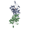
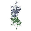
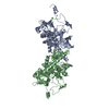
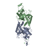
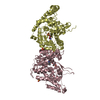
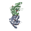
| Title | Crystal structure of Glu335Gln mutant of Clostridium botulinum neurotoxin E catalytic domain | ||||||
 Components Components | botulinum neurotoxin type E | ||||||
 Keywords Keywords | HYDROLASE / botulinum neurotoxin E / catalytic domain / light chain / Glu335Gln mutant / apoenzyme | ||||||
| Function / homology |  Function and homology information Function and homology informationToxicity of botulinum toxin type E (botE) / bontoxilysin / host cell presynaptic membrane / host cell cytoplasmic vesicle / host cell cytosol / protein transmembrane transporter activity / metalloendopeptidase activity / toxin activity / lipid binding / host cell plasma membrane ...Toxicity of botulinum toxin type E (botE) / bontoxilysin / host cell presynaptic membrane / host cell cytoplasmic vesicle / host cell cytosol / protein transmembrane transporter activity / metalloendopeptidase activity / toxin activity / lipid binding / host cell plasma membrane / proteolysis / extracellular region / zinc ion binding / membrane Similarity search - Function | ||||||
| Biological species |   Clostridium botulinum (bacteria) Clostridium botulinum (bacteria) | ||||||
| Method |  X-RAY DIFFRACTION / SYNCHROTRON / FOURIER SYNTHESIS / Resolution: 2.6 Å X-RAY DIFFRACTION / SYNCHROTRON / FOURIER SYNTHESIS / Resolution: 2.6 Å | ||||||
 Authors Authors | Agarwal, R. / Binz, T. / Swaminathan, S. | ||||||
 Citation Citation | Journal: Biochemistry / Year: 2005 Title: Analysis of Active Site Residues of Botulinum Neurotoxin E by Mutational, Functional, and Structural Studies: Glu335Gln Is an Apoenzyme. Authors: Agarwal, R. / Binz, T. / Swaminathan, S. | ||||||
| History |
|
- Structure visualization
| Structure viewer | Molecule: MolmilJmol/JSmol |
|---|
- Downloads & links
-Download
| PDBx/mmCIF format | 1zl5.cif.gz | 170.8 KB | Display | PDBx/mmCIF format |
|---|---|---|---|---|
| PDB format | pdb1zl5.ent.gz | 135.4 KB | Display | PDB format |
| PDBx/mmJSON format | 1zl5.json.gz | Tree view | PDBx/mmJSON format | |
| Others |  Other downloads Other downloads |
-Validation report
| Summary document | 1zl5_validation.pdf.gz | 439.7 KB | Display | wwPDB validaton report |
|---|---|---|---|---|
| Full document | 1zl5_full_validation.pdf.gz | 458.3 KB | Display | |
| Data in XML | 1zl5_validation.xml.gz | 32.8 KB | Display | |
| Data in CIF | 1zl5_validation.cif.gz | 45.8 KB | Display | |
| Arichive directory | https://data.pdbj.org/pub/pdb/validation_reports/zl/1zl5ftp://data.pdbj.org/pub/pdb/validation_reports/zl/1zl5 | HTTPS FTP |
-Related structure data
| Related structure data |  1zkwC  1zkxC  1zl6C  1zn3C  1t3aS S: Starting model for refinement C: citing same article ( |
|---|---|
| Similar structure data |


-Links
 PDBj
PDBj
- Assembly
| Deposited unit | 
| ||||||||
|---|---|---|---|---|---|---|---|---|---|
| 1 |
| ||||||||
| 2 | 
| ||||||||
| Unit cell |
|
-Components
| #1: Protein | Mass: 47686.797 Da / Num. of mol.: 2 / Fragment: catalytic domain (residues 2-421) / Mutation: E335Q Source method: isolated from a genetically manipulated source Source: (gene. exp.) Clostridium botulinum (bacteria) / Strain: NCTC-11219 / Plasmid: PET-9C / Species (production host): Escherichia coli / Production host: #2: Chemical | ChemComp-CL /   Mass: 35.453 Da / Num. of mol.: 8 / Source method: obtained synthetically / Formula: Cl Mass: 35.453 Da / Num. of mol.: 8 / Source method: obtained synthetically / Formula: Cl#3: Water | ChemComp-HOH / |  Mass: 18.015 Da / Num. of mol.: 250 / Source method: isolated from a natural source / Formula: H2O Mass: 18.015 Da / Num. of mol.: 250 / Source method: isolated from a natural source / Formula: H2O |
|---|
-Experimental details
-Experiment
| Experiment | Method: X-RAY DIFFRACTION / Number of used crystals: 1 |
|---|
- Sample preparation
| Crystal | Density Matthews: 2.67 Å3/Da / Density % sol: 48 % |
|---|---|
| Crystal grow | Temperature: 298 K / Method: vapor diffusion, sitting drop / pH: 5.6 Details: ammonium sulfate, lithium sulfate, sodium citrate, pH 5.6, VAPOR DIFFUSION, SITTING DROP, temperature 298K |
-Data collection
| Diffraction | Mean temperature: 100 K |
|---|---|
| Diffraction source | Source: SYNCHROTRON / Site: NSLS  / Beamline: X29A / Wavelength: 1.1 Å / Beamline: X29A / Wavelength: 1.1 Å |
| Detector | Type: ADSC QUANTUM 315 / Detector: CCD / Date: Jun 2, 2004 / Details: mirrors |
| Radiation | Monochromator: graphite / Protocol: SINGLE WAVELENGTH / Monochromatic (M) / Laue (L): M / Scattering type: x-ray |
| Radiation wavelength | Wavelength: 1.1 Å / Relative weight: 1 |
| Reflection | Resolution: 2.6→50 Å / Num. obs: 30978 / % possible obs: 89.8 % / Observed criterion σ(F): 0 / Observed criterion σ(I): 0 / Redundancy: 6.9 % / Biso Wilson estimate: 13.4 Å2 / Rmerge(I) obs: 0.12 / Net I/σ(I): 4.8 |
| Reflection shell | Resolution: 2.6→2.69 Å / Redundancy: 3.4 % / Rmerge(I) obs: 0.44 / Mean I/σ(I) obs: 2 / Num. unique all: 1878 / % possible all: 55.3 |
- Processing
| Software |
| ||||||||||||||||||||||||||||||||||||
|---|---|---|---|---|---|---|---|---|---|---|---|---|---|---|---|---|---|---|---|---|---|---|---|---|---|---|---|---|---|---|---|---|---|---|---|---|---|
| Refinement | Method to determine structure: FOURIER SYNTHESIS Starting model: PDB entry 1T3A Resolution: 2.6→46.55 Å / Rfactor Rfree error: 0.013 / Data cutoff high absF: 47375.49 / Data cutoff low absF: 0 / Isotropic thermal model: RESTRAINED / Cross valid method: THROUGHOUT / σ(F): 0 Details: In general, three loop regions (50-65, 188-200 and 234-244) are either disordered or have very weak electron density in both chains. In this entry, the missing residues are due to the ...Details: In general, three loop regions (50-65, 188-200 and 234-244) are either disordered or have very weak electron density in both chains. In this entry, the missing residues are due to the absence of interpretable electron density in those regions.
| ||||||||||||||||||||||||||||||||||||
| Solvent computation | Solvent model: FLAT MODEL / Bsol: 22.9893 Å2 / ksol: 0.330847 e/Å3 | ||||||||||||||||||||||||||||||||||||
| Displacement parameters | Biso mean: 37.1 Å2
| ||||||||||||||||||||||||||||||||||||
| Refine analyze |
| ||||||||||||||||||||||||||||||||||||
| Refinement step | Cycle: LAST / Resolution: 2.6→46.55 Å
| ||||||||||||||||||||||||||||||||||||
| Refine LS restraints |
| ||||||||||||||||||||||||||||||||||||
| LS refinement shell | Resolution: 2.6→2.76 Å / Rfactor Rfree error: 0.077 / Total num. of bins used: 6
| ||||||||||||||||||||||||||||||||||||
| Xplor file |
|
