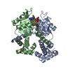
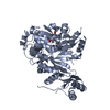
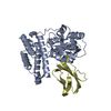
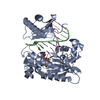
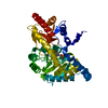
+Open data
-Basic information
Entry | Database: PDB / ID: 1zh1 | ||||||
---|---|---|---|---|---|---|---|
Title | Structure of the zinc-binding domain of HCV NS5A | ||||||
Components | non-structural polyprotein | ||||||
Keywords | METAL BINDING PROTEIN / HCV / nonstructural protein 5A / NS5A / Domain I | ||||||
Function / homology | Function and homology information positive stranded viral RNA replication / hepacivirin / host cell mitochondrial membrane / host cell lipid droplet / symbiont-mediated suppression of host TRAF-mediated signal transduction / transformation of host cell by virus / symbiont-mediated perturbation of host cell cycle G1/S transition checkpoint / symbiont-mediated suppression of host JAK-STAT cascade via inhibition of STAT1 activity / symbiont-mediated suppression of host cytoplasmic pattern recognition receptor signaling pathway via inhibition of MAVS activity / viral genome replication ...positive stranded viral RNA replication / hepacivirin / host cell mitochondrial membrane / host cell lipid droplet / symbiont-mediated suppression of host TRAF-mediated signal transduction / transformation of host cell by virus / symbiont-mediated perturbation of host cell cycle G1/S transition checkpoint / symbiont-mediated suppression of host JAK-STAT cascade via inhibition of STAT1 activity / symbiont-mediated suppression of host cytoplasmic pattern recognition receptor signaling pathway via inhibition of MAVS activity / viral genome replication / SH3 domain binding / nucleoside-triphosphate phosphatase / protein complex oligomerization / monoatomic ion channel activity / viral nucleocapsid / clathrin-dependent endocytosis of virus by host cell / Hydrolases; Acting on peptide bonds (peptidases); Cysteine endopeptidases / RNA helicase activity / host cell perinuclear region of cytoplasm / host cell endoplasmic reticulum membrane / symbiont-mediated suppression of host type I interferon-mediated signaling pathway / RNA helicase / ribonucleoprotein complex / induction by virus of host autophagy / serine-type endopeptidase activity / cysteine-type endopeptidase activity / RNA-directed RNA polymerase / viral RNA genome replication / virus-mediated perturbation of host defense response / RNA-dependent RNA polymerase activity / fusion of virus membrane with host endosome membrane / viral envelope / host cell nucleus / virion attachment to host cell / apoptotic process / host cell plasma membrane / structural molecule activity / virion membrane / ATP hydrolysis activity / protein-containing complex / proteolysis / RNA binding / zinc ion binding / ATP binding / identical protein binding / membrane Similarity search - Function | ||||||
Biological species | Hepatitis C virus | ||||||
Method | X-RAY DIFFRACTION / SYNCHROTRON / MAD / Resolution: 2.5 Å | ||||||
Authors | Tellinghuisen, T.L. / Marcotrigiano, J. / Rice, C.M. | ||||||
Citation | Journal: Nature / Year: 2005 Title: Structure of the zinc-binding domain of an essential component of the hepatitis C virus replicase. Authors: Tellinghuisen, T.L. / Marcotrigiano, J. / Rice, C.M. | ||||||
History |
|
-Structure visualization
Structure viewer | Molecule: MolmilJmol/JSmol |
---|
-Downloads & links
-Download
PDBx/mmCIF format | 1zh1.cif.gz | 71.6 KB | Display | PDBx/mmCIF format |
---|---|---|---|---|
PDB format | pdb1zh1.ent.gz | 57.4 KB | Display | PDB format |
PDBx/mmJSON format | 1zh1.json.gz | Tree view | PDBx/mmJSON format | |
Others | Other downloads |
-Validation report
Summary document | 1zh1_validation.pdf.gz | 432.8 KB | Display | wwPDB validaton report |
---|---|---|---|---|
Full document | 1zh1_full_validation.pdf.gz | 443.5 KB | Display | |
Data in XML | 1zh1_validation.xml.gz | 15.9 KB | Display | |
Data in CIF | 1zh1_validation.cif.gz | 21.4 KB | Display | |
Arichive directory | https://data.pdbj.org/pub/pdb/validation_reports/zh/1zh1 ftp://data.pdbj.org/pub/pdb/validation_reports/zh/1zh1 | HTTPS FTP |
-Related structure data
Similar structure data |
---|
-Links
-Assembly
Deposited unit |
| ||||||||
---|---|---|---|---|---|---|---|---|---|
1 |
| ||||||||
Unit cell |
|
-Components
#1: Protein | Mass: 18008.484 Da / Num. of mol.: 2 / Fragment: Domain I; zinc binding domain Source method: isolated from a genetically manipulated source Source: (gene. exp.) Hepatitis C virus / Genus: Hepacivirus / Gene: Nonstructural protein 5A / Plasmid: pET30b / Species (production host): Escherichia coli / Production host: Escherichia coli BL21(DE3) (bacteria) / Strain (production host): BL21(de3) / References: UniProt: Q9WMX2 #2: Chemical | #3: Water | ChemComp-HOH / | |
---|
-Experimental details
-Experiment
Experiment | Method: X-RAY DIFFRACTION / Number of used crystals: 1 |
---|
-Sample preparation
Crystal | Density Matthews: 2.7 Å3/Da / Density % sol: 54.5 % | ||||||||||||||||||||||||||||||||||||||||||||||||
---|---|---|---|---|---|---|---|---|---|---|---|---|---|---|---|---|---|---|---|---|---|---|---|---|---|---|---|---|---|---|---|---|---|---|---|---|---|---|---|---|---|---|---|---|---|---|---|---|---|
Crystal grow | Temperature: 277 K / Method: vapor diffusion, hanging drop / pH: 7 Details: isopropanol, trisodium citrate, non-detergent sulphobetaine 201, HEPES, pH 7.0, VAPOR DIFFUSION, HANGING DROP, temperature 277K | ||||||||||||||||||||||||||||||||||||||||||||||||
Crystal grow | *PLUS Temperature: 4 ℃ / pH: 8 | ||||||||||||||||||||||||||||||||||||||||||||||||
Components of the solutions | *PLUS
|
-Data collection
Diffraction | Mean temperature: 100 K | ||||||||||||
---|---|---|---|---|---|---|---|---|---|---|---|---|---|
Diffraction source | Source: SYNCHROTRON / Site: NSLS / Beamline: X9A / Wavelength: 1.27069, 1.28385, 1.28345 | ||||||||||||
Detector | Type: MARRESEARCH / Detector: CCD / Date: Sep 14, 2004 | ||||||||||||
Radiation | Monochromator: Si(111) / Protocol: MAD / Monochromatic (M) / Laue (L): M / Scattering type: x-ray | ||||||||||||
Radiation wavelength |
| ||||||||||||
Reflection | Resolution: 2.5→30 Å / Num. obs: 18007 / % possible obs: 100 % / Observed criterion σ(I): 4 / Redundancy: 5 % / Rsym value: 0.087 / Net I/σ(I): 35.6 | ||||||||||||
Reflection shell | Resolution: 2.5→2.59 Å / Redundancy: 5 % / Rmerge(I) obs: 0.274 / Mean I/σ(I) obs: 5.7 / % possible all: 100 | ||||||||||||
Reflection | *PLUS Highest resolution: 2.5 Å / Lowest resolution: 30 Å / % possible obs: 100 % / Num. measured all: 314341 / Rmerge(I) obs: 0.087 | ||||||||||||
Reflection shell | *PLUS % possible obs: 100 % |
-Processing
Software |
| ||||||||||||||||||||
---|---|---|---|---|---|---|---|---|---|---|---|---|---|---|---|---|---|---|---|---|---|
Refinement | Method to determine structure: MAD / Resolution: 2.5→30 Å / σ(F): 2 / Stereochemistry target values: Engh & Huber / Details: The reflection file includes anomalous pairs.
| ||||||||||||||||||||
Refinement step | Cycle: LAST / Resolution: 2.5→30 Å
| ||||||||||||||||||||
Refine LS restraints |
| ||||||||||||||||||||
Software | *PLUS Name: CNS / Version: 1 / Classification: refinement | ||||||||||||||||||||
Refinement | *PLUS Highest resolution: 2.5 Å / Lowest resolution: 30 Å / σ(F): 2 / % reflection Rfree: 10 % / Rfactor obs: 0.225 | ||||||||||||||||||||
Solvent computation | *PLUS | ||||||||||||||||||||
Displacement parameters | *PLUS | ||||||||||||||||||||
Refine LS restraints | *PLUS
|