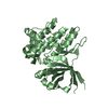
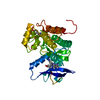
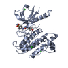
Entry Database : PDB / ID : 1zgyTitle Structural and Biochemical Basis for Selective Repression of the Orphan Nuclear Receptor LRH-1 by SHP Nuclear receptor subfamily 0, group B, member 2 Peroxisome proliferator activated receptor gamma Keywords / Function / homology Function Domain/homology Component
/ / / / / / / / / / / / / / / / / / / / / / / / / / / / / / / / / / / / / / / / / / / / / / / / / / / / / / / / / / / / / / / / / / / / / / / / / / / / / / / / / / / / / / / / / / / / / / / / / / / / / / / / / / / / / / / / / / / / / / / / / / / / / / / / / / / / / / / / / / Biological species Homo sapiens (human)Rattus norvegicus (Norway rat)Method / / / Resolution : 1.8 Å Authors Li, Y. / Choi, M. / Suino, K. / Kovach, A. / Daugherty, J. / Kliewer, S.A. / Xu, H.E. Journal : Proc.Natl.Acad.Sci.Usa / Year : 2005Title : Structural and biochemical basis for selective repression of the orphan nuclear receptor liver receptor homolog 1 by small heterodimer partner.Authors : Li, Y. / Choi, M. / Suino, K. / Kovach, A. / Daugherty, J. / Kliewer, S.A. / Xu, H.E. History Deposition Apr 22, 2005 Deposition site / Processing site Revision 1.0 Jul 26, 2005 Provider / Type Revision 1.1 Apr 30, 2008 Group Revision 1.2 Jul 13, 2011 Group Revision 1.3 Oct 11, 2017 Group / Category / Item / _software.nameRevision 1.4 Aug 23, 2023 Group Data collection / Database references ... Data collection / Database references / Derived calculations / Refinement description Category chem_comp_atom / chem_comp_bond ... chem_comp_atom / chem_comp_bond / database_2 / pdbx_initial_refinement_model / struct_site Item _database_2.pdbx_DOI / _database_2.pdbx_database_accession ... _database_2.pdbx_DOI / _database_2.pdbx_database_accession / _struct_site.pdbx_auth_asym_id / _struct_site.pdbx_auth_comp_id / _struct_site.pdbx_auth_seq_id
Show all Show less
 Movie
Movie Controller

 Yorodumi
 Open data
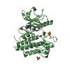
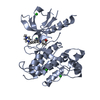
 Basic information
 Components
 Keywords
 Function and homology information
 Homo sapiens (human)
 X-RAY DIFFRACTION /
  Authors
 Citation
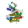
 Structure visualization
 Downloads & links
 Other downloads

 PDBj
PDBj

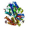
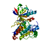
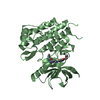
 Assembly
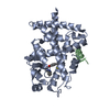
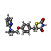
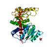
 Mass: 357.427 Da / Num. of mol.: 1 / Source method: obtained synthetically / Formula: C18H19N3O3S
 Mass: 18.015 Da / Num. of mol.: 158 / Source method: isolated from a natural source / Formula: H2O
 Sample preparation
 / Beamline: 32-ID / Wavelength: 0.99998 Å
 Processing