- PDB-1zaw: Ribosomal Protein L10-L12(NTD) Complex, Space Group P212121, Form A -
+
Open data
ID or keywords:
Loading...
-
Basic information
Entry
Database: PDB / ID: 1zaw
Title
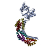
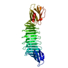
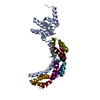
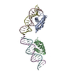
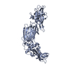
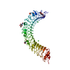
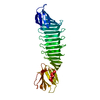
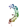
Ribosomal Protein L10-L12(NTD) Complex, Space Group P212121, Form A
Components
50S ribosomal protein L10
50S ribosomal protein L7/L12
Keywords
STRUCTURAL PROTEIN / ribosome structure and function / L10-L12 complex structure / L10E structure / L7/12 ribosomal stalk / thiostrepton loop of 23S rRNA / translation factor recruitment / GTPase stimulation / mechanism of translation / rapid kinetics
Function / homology
Function and homology information
large ribosomal subunit rRNA binding / cytosolic large ribosomal subunit / structural constituent of ribosome / translation / mRNA binding Similarity search - Function
Single alpha-helices involved in coiled-coils or other helix-helix interfaces - #290 / Ribosomal protein uL10 C-terminal region / Ribosomal protein L10, N-terminal RNA-binding domain / Ribosomal protein L7/L12, oligomerisation / Ribosomal protein L7/L12, oligomerisation domain superfamily / Ribosomal protein L7/L12 dimerisation domain / Ribosomal protein L7/L12 / Ribosomal protein L7/L12, C-terminal / Ribosomal protein L7/L12 C-terminal domain / Ribosomal protein L7/L12, C-terminal/adaptor protein ClpS-like ...Single alpha-helices involved in coiled-coils or other helix-helix interfaces - #290 / Ribosomal protein uL10 C-terminal region / Ribosomal protein L10, N-terminal RNA-binding domain / Ribosomal protein L7/L12, oligomerisation / Ribosomal protein L7/L12, oligomerisation domain superfamily / Ribosomal protein L7/L12 dimerisation domain / Ribosomal protein L7/L12 / Ribosomal protein L7/L12, C-terminal / Ribosomal protein L7/L12 C-terminal domain / Ribosomal protein L7/L12, C-terminal/adaptor protein ClpS-like / Ribosomal protein L10, eubacterial, conserved site / Ribosomal protein L10 signature. / Ribosomal protein L10 / Single alpha-helices involved in coiled-coils or other helix-helix interfaces / : / Ribosomal protein L10-like domain superfamily / Helix non-globular / Ribosomal protein L10P / Ribosomal protein L10 / Special / Alpha-Beta Plaits / 2-Layer Sandwich / Alpha Beta Similarity search - Domain/homology
Journal: Cell / Year: 2005 Title: Structural basis for the function of the ribosomal L7/12 stalk in factor binding and GTPase activation. Authors: Mihaela Diaconu / Ute Kothe / Frank Schlünzen / Niels Fischer / Jörg M Harms / Alexander G Tonevitsky / Holger Stark / Marina V Rodnina / Markus C Wahl / Abstract: The L7/12 stalk of the large subunit of bacterial ribosomes encompasses protein L10 and multiple copies of L7/12. We present crystal structures of Thermotoga maritima L10 in complex with three L7/12 ...The L7/12 stalk of the large subunit of bacterial ribosomes encompasses protein L10 and multiple copies of L7/12. We present crystal structures of Thermotoga maritima L10 in complex with three L7/12 N-terminal-domain dimers, refine the structure of an archaeal L10E N-terminal domain on the 50S subunit, and identify these elements in cryo-electron-microscopic reconstructions of Escherichia coli ribosomes. The mobile C-terminal helix alpha8 of L10 carries three L7/12 dimers in T. maritima and two in E. coli, in concordance with the different length of helix alpha8 of L10 in these organisms. The stalk is organized into three elements (stalk base, L10 helix alpha8-L7/12 N-terminal-domain complex, and L7/12 C-terminal domains) linked by flexible connections. Highly mobile L7/12 C-terminal domains promote recruitment of translation factors to the ribosome and stimulate GTP hydrolysis by the ribosome bound factors through stabilization of their active GTPase conformation.
A: 50S ribosomal protein L10 U: 50S ribosomal protein L7/L12 V: 50S ribosomal protein L7/L12 W: 50S ribosomal protein L7/L12 X: 50S ribosomal protein L7/L12 Y: 50S ribosomal protein L7/L12 Z: 50S ribosomal protein L7/L12
In the structure databanks used in Yorodumi, some data are registered as the other names, "COVID-19 virus" and "2019-nCoV". Here are the details of the virus and the list of structure data.
Jan 31, 2019. EMDB accession codes are about to change! (news from PDBe EMDB page)
EMDB accession codes are about to change! (news from PDBe EMDB page)
The allocation of 4 digits for EMDB accession codes will soon come to an end. Whilst these codes will remain in use, new EMDB accession codes will include an additional digit and will expand incrementally as the available range of codes is exhausted. The current 4-digit format prefixed with “EMD-” (i.e. EMD-XXXX) will advance to a 5-digit format (i.e. EMD-XXXXX), and so on. It is currently estimated that the 4-digit codes will be depleted around Spring 2019, at which point the 5-digit format will come into force.
The EM Navigator/Yorodumi systems omit the EMD- prefix.
Related info.:Q: What is EMD? / ID/Accession-code notation in Yorodumi/EM Navigator
Yorodumi is a browser for structure data from EMDB, PDB, SASBDB, etc.
This page is also the successor to EM Navigator detail page, and also detail information page/front-end page for Omokage search.
The word "yorodu" (or yorozu) is an old Japanese word meaning "ten thousand". "mi" (miru) is to see.
Related info.:EMDB / PDB / SASBDB / Comparison of 3 databanks / Yorodumi Search / Aug 31, 2016. New EM Navigator & Yorodumi / Yorodumi Papers / Jmol/JSmol / Function and homology information / Changes in new EM Navigator and Yorodumi
 Movie
Movie Controller

 Yorodumi
 Open data
 Basic information
 Components
 Keywords
 Function and homology information
 Thermotoga maritima (bacteria)
 X-RAY DIFFRACTION /
  Authors
 Citation
 Structure visualization
 Downloads & links
 Other downloads
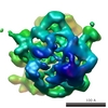


 PDBj
PDBj


 Assembly
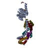
 Mass: 18.015 Da / Num. of mol.: 223 / Source method: isolated from a natural source / Formula: H2O
 Sample preparation
 Processing