 Movie
Movie Controller

[English] 日本語
 Yorodumi
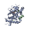
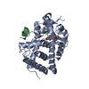
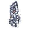
- PDB-1zak: ADENYLATE KINASE FROM MAIZE IN COMPLEX WITH THE INHIBITOR P1,P5-B... -
+ Open data

- Basic information
| Entry | Database: PDB / ID: 1zak | ||||||
|---|---|---|---|---|---|---|---|
| Title | ADENYLATE KINASE FROM MAIZE IN COMPLEX WITH THE INHIBITOR P1,P5-BIS(ADENOSINE-5'-)PENTAPHOSPHATE (AP5A) | ||||||
 Components Components | ADENYLATE KINASE | ||||||
 Keywords Keywords | TRANSFERASE / ATP:AMP-PHOSPHOTRANSFERASE | ||||||
| Function / homology |  Function and homology information Function and homology informationadenylate kinase / AMP kinase activity / chloroplast / ATP binding / cytoplasm Similarity search - Function | ||||||
| Biological species |  | ||||||
| Method |  X-RAY DIFFRACTION / MOLECULAR REPLACEMENT / Resolution: 3.5 Å X-RAY DIFFRACTION / MOLECULAR REPLACEMENT / Resolution: 3.5 Å | ||||||
 Authors Authors | Wild, K. / Schulz, G.E. | ||||||
 Citation Citation | Journal: Eur.J.Biochem. / Year: 1997 Title: Structure, catalysis and supramolecular assembly of adenylate kinase from maize. Authors: Wild, K. / Grafmuller, R. / Wagner, E. / Schulz, G.E. #1: Journal: Eur.J.Biochem. / Year: 1994Title: Primary Structure of Maize Chloroplast Adenylate Kinase Authors: Schiltz, E. / Burger, S. / Grafmuller, R. / Deppert, W.R. / Haehnel, W. / Wagner, E. | ||||||
| History |
|
- Structure visualization
| Structure viewer | Molecule: MolmilJmol/JSmol |
|---|
- Downloads & links
-Download
| PDBx/mmCIF format | 1zak.cif.gz | 88.2 KB | Display | PDBx/mmCIF format |
|---|---|---|---|---|
| PDB format | pdb1zak.ent.gz | 66.4 KB | Display | PDB format |
| PDBx/mmJSON format | 1zak.json.gz | Tree view | PDBx/mmJSON format | |
| Others |  Other downloads Other downloads |
-Validation report
| Arichive directory | https://data.pdbj.org/pub/pdb/validation_reports/za/1zakftp://data.pdbj.org/pub/pdb/validation_reports/za/1zak | HTTPS FTP |
|---|
-Related structure data
| Related structure data | 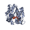 1akeS S: Starting model for refinement |
|---|---|
| Similar structure data |


-Links
 PDBj
PDBj- Assembly
| Deposited unit | 
| ||||||||
|---|---|---|---|---|---|---|---|---|---|
| 1 |
| ||||||||
| Unit cell |
| ||||||||
| Noncrystallographic symmetry (NCS) | NCS oper: (Code: given Matrix: (-0.7065, 0.7077, 0.0046), Vector: |
-Components
| #1: Protein | Mass: 24897.385 Da / Num. of mol.: 2 / Source method: isolated from a natural source / Source: (natural) #2: Chemical | 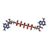  Mass: 916.367 Da / Num. of mol.: 2 / Source method: obtained synthetically / Formula: C20H29N10O22P5 Mass: 916.367 Da / Num. of mol.: 2 / Source method: obtained synthetically / Formula: C20H29N10O22P5Nonpolymer details | THE WATER MOLECULE MODELED INTO THE AMP-BINDING SITE TOGETHER WITH CMP HAS THE COORDINATES X Y Z 17. ...THE WATER MOLECULE MODELED INTO THE AMP-BINDING SITE TOGETHER WITH CMP HAS THE COORDINATE | |
|---|
-Experimental details
-Experiment
| Experiment | Method: X-RAY DIFFRACTION / Number of used crystals: 1 |
|---|
- Sample preparation
| Crystal | Density Matthews: 2.8 Å3/Da / Density % sol: 56 % | ||||||||||||||||||||||||||||||||||||||||||||||||
|---|---|---|---|---|---|---|---|---|---|---|---|---|---|---|---|---|---|---|---|---|---|---|---|---|---|---|---|---|---|---|---|---|---|---|---|---|---|---|---|---|---|---|---|---|---|---|---|---|---|
| Crystal grow | pH: 7.5 / Details: pH 7.5 | ||||||||||||||||||||||||||||||||||||||||||||||||
| Crystal grow | *PLUS Method: vapor diffusion, hanging drop | ||||||||||||||||||||||||||||||||||||||||||||||||
| Components of the solutions | *PLUS
|
-Data collection
| Diffraction | Mean temperature: 293 K |
|---|---|
| Diffraction source | Source: ROTATING ANODE / Type: RIGAKU RUH2R / Wavelength: 1.5418 |
| Detector | Type: SIEMENS-NICOLET X100 / Detector: AREA DETECTOR / Date: Apr 1, 1994 |
| Radiation | Monochromator: GRAPHITE(002) / Monochromatic (M) / Laue (L): M / Scattering type: x-ray |
| Radiation wavelength | Wavelength: 1.5418 Å / Relative weight: 1 |
| Reflection | Resolution: 3.16→100 Å / Num. obs: 6998 / % possible obs: 75.4 % / Redundancy: 3 % / Rsym value: 0.074 |
| Reflection shell | Resolution: 3.16→3.33 Å / Rsym value: 0.172 / % possible all: 53.3 |
| Reflection | *PLUS Rmerge(I) obs: 0.074 |
- Processing
| Software |
| ||||||||||||||||||||||||||||||||||||||||||||||||||||||||||||
|---|---|---|---|---|---|---|---|---|---|---|---|---|---|---|---|---|---|---|---|---|---|---|---|---|---|---|---|---|---|---|---|---|---|---|---|---|---|---|---|---|---|---|---|---|---|---|---|---|---|---|---|---|---|---|---|---|---|---|---|---|---|
| Refinement | Method to determine structure: MOLECULAR REPLACEMENT Starting model: PDB ENTRY 1AKE Resolution: 3.5→25 Å / Details: ONLY THE OVERALL B-FACTOR WAS REFINED.
| ||||||||||||||||||||||||||||||||||||||||||||||||||||||||||||
| Displacement parameters | Biso mean: 23.8 Å2 | ||||||||||||||||||||||||||||||||||||||||||||||||||||||||||||
| Refinement step | Cycle: LAST / Resolution: 3.5→25 Å
| ||||||||||||||||||||||||||||||||||||||||||||||||||||||||||||
| Refine LS restraints |
| ||||||||||||||||||||||||||||||||||||||||||||||||||||||||||||
| Refine LS restraints NCS | NCS model details: CONSTRAINED | ||||||||||||||||||||||||||||||||||||||||||||||||||||||||||||
| LS refinement shell | Resolution: 3.5→3.66 Å / Total num. of bins used: 8
| ||||||||||||||||||||||||||||||||||||||||||||||||||||||||||||
| Xplor file |
| ||||||||||||||||||||||||||||||||||||||||||||||||||||||||||||
| Software | *PLUS Name: X-PLOR / Version: 3.1 / Classification: refinement | ||||||||||||||||||||||||||||||||||||||||||||||||||||||||||||
| Refine LS restraints | *PLUS
|
