 Movie
Movie Controller

[English] 日本語
 Yorodumi
- PDB-1za6: The structure of an antitumor CH2-domain-deleted humanized antibody -
+ Open data

- Basic information
| Entry | Database: PDB / ID: 1za6 | ||||||
|---|---|---|---|---|---|---|---|
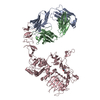
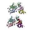
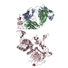
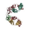
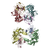
| Title | The structure of an antitumor CH2-domain-deleted humanized antibody | ||||||
 Components Components |
| ||||||
 Keywords Keywords | IMMUNE SYSTEM / Immunoglobulin fold / CH2-domain-deletion | ||||||
| Function / homology |  Function and homology information Function and homology informationimmunoglobulin complex / adaptive immune response / extracellular region / plasma membrane Similarity search - Function | ||||||
| Biological species |  Homo sapiens (human) Homo sapiens (human) | ||||||
| Method |  X-RAY DIFFRACTION / SYNCHROTRON / MOLECULAR REPLACEMENT / Resolution: 2.8 Å X-RAY DIFFRACTION / SYNCHROTRON / MOLECULAR REPLACEMENT / Resolution: 2.8 Å | ||||||
 Authors Authors | Larson, S.B. / Day, J.S. / Glaser, S. / Braslawsky, G. / McPherson, A. | ||||||
 Citation Citation | Journal: J.Mol.Biol. / Year: 2005 Title: The Structure of an Antitumor C(H)2-domain-deleted Humanized Antibody. Authors: Larson, S.B. / Day, J.S. / Glaser, S. / Braslawsky, G. / McPherson, A. #1: Journal: Acta Crystallogr.,Sect.D / Year: 2005 Title: Combined use of AFM and X-ray diffraction to analyze crystals of an engineered, domain-deleted antibody Authors: Larson, S.B. / Kuznetsov, Y.G. / Day, J. / Zhou, J. / Glaser, S. / Braslawsky, G. / McPherson, A. | ||||||
| History |
| ||||||
| Remark 999 | SEQUENCE There was no suitable sequence database match at time of processing. |
- Structure visualization
| Structure viewer | Molecule: MolmilJmol/JSmol |
|---|
- Downloads & links
-Download
| PDBx/mmCIF format | 1za6.cif.gz | 411.8 KB | Display | PDBx/mmCIF format |
|---|---|---|---|---|
| PDB format | pdb1za6.ent.gz | 340.3 KB | Display | PDB format |
| PDBx/mmJSON format | 1za6.json.gz | Tree view | PDBx/mmJSON format | |
| Others |  Other downloads Other downloads |
-Validation report
| Arichive directory | https://data.pdbj.org/pub/pdb/validation_reports/za/1za6ftp://data.pdbj.org/pub/pdb/validation_reports/za/1za6 | HTTPS FTP |
|---|
-Related structure data


-Links
 PDBj
PDBj

- Assembly
| Deposited unit | 
| |||||||||||||||||||||||||||
|---|---|---|---|---|---|---|---|---|---|---|---|---|---|---|---|---|---|---|---|---|---|---|---|---|---|---|---|---|
| 1 | 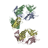
| |||||||||||||||||||||||||||
| 2 | 
| |||||||||||||||||||||||||||
| 3 | 
| |||||||||||||||||||||||||||
| 4 |
| |||||||||||||||||||||||||||
| Unit cell |
| |||||||||||||||||||||||||||
| Noncrystallographic symmetry (NCS) | NCS domain:
| |||||||||||||||||||||||||||
| Details | Chains A, B, C, D are the presumed biological unit / Chains E, F, G, H are the presumed biological unit |
-Components
| #1: Antibody | Mass: 24206.863 Da / Num. of mol.: 4 Source method: isolated from a genetically manipulated source Source: (gene. exp.) Homo sapiens (human) / Production host:   Cricetulus griseus (Chinese hamster) / References: UniProt: Q6GMV9 Cricetulus griseus (Chinese hamster) / References: UniProt: Q6GMV9#2: Antibody | Mass: 36952.125 Da / Num. of mol.: 4 Source method: isolated from a genetically manipulated source Source: (gene. exp.) Homo sapiens (human) / Production host: Cricetulus griseus (Chinese hamster) / References: UniProt: Q6GMX6Has protein modification | Y | |
|---|
-Experimental details
-Experiment
| Experiment | Method: X-RAY DIFFRACTION / Number of used crystals: 1 |
|---|
- Sample preparation
| Crystal | Density Matthews: 3.29 Å3/Da / Density % sol: 62.3 % |
|---|---|
| Crystal grow | Temperature: 290 K / Method: vapor diffusion, sitting drop / pH: 7.2 Details: 4 M sodium formate, 1.5 mM Triton X-100 detergent, pH 7.2, VAPOR DIFFUSION, SITTING DROP, temperature 290K, pH 7.20 |
-Data collection
| Diffraction | Mean temperature: 100 K |
|---|---|
| Diffraction source | Source: SYNCHROTRON / Site: ALS  / Beamline: 5.0.2 / Wavelength: 0.9194 / Beamline: 5.0.2 / Wavelength: 0.9194 |
| Detector | Type: ADSC QUANTUM 4 / Detector: CCD / Date: Oct 4, 2003 |
| Radiation | Monochromator: DOUBLE CRYSTAL, SI(1 1 1) / Protocol: SINGLE WAVELENGTH / Monochromatic (M) / Laue (L): M / Scattering type: x-ray |
| Radiation wavelength | Wavelength: 0.9194 Å / Relative weight: 1 |
| Reflection | Resolution: 2.8→45.13 Å / Num. obs: 76662 / % possible obs: 99.9 % / Observed criterion σ(I): 0 / Redundancy: 9.06 % / Biso Wilson estimate: 22.7 Å2 / Rmerge(I) obs: 0.152 / Net I/σ(I): 10.9 |
| Reflection shell | Resolution: 2.8→2.9 Å / Redundancy: 7.62 % / Rmerge(I) obs: 0.365 / Mean I/σ(I) obs: 5 / % possible all: 98 |
- Processing
| Software |
| ||||||||||||||||||||||||||||||||||||||||||||||||||||||||||||||||||||||||||||||||
|---|---|---|---|---|---|---|---|---|---|---|---|---|---|---|---|---|---|---|---|---|---|---|---|---|---|---|---|---|---|---|---|---|---|---|---|---|---|---|---|---|---|---|---|---|---|---|---|---|---|---|---|---|---|---|---|---|---|---|---|---|---|---|---|---|---|---|---|---|---|---|---|---|---|---|---|---|---|---|---|---|---|
| Refinement | Method to determine structure: MOLECULAR REPLACEMENT Starting model: PDB ENTRY 1BBJ; PDB ENTRY 1FC1 Resolution: 2.8→45.13 Å / Rfactor Rfree error: 0.004 / Data cutoff high absF: 38542.32 / Data cutoff low absF: 0 / Isotropic thermal model: RESTRAINED / Cross valid method: THROUGHOUT / σ(F): 4 / Stereochemistry target values: ENGH & HUBER / Details: USED NCS RESTRAINTS FOR EQUIVALENT CHAINS
| ||||||||||||||||||||||||||||||||||||||||||||||||||||||||||||||||||||||||||||||||
| Solvent computation | Solvent model: FLAT MODEL / Bsol: 29.13 Å2 / ksol: 0.33 e/Å3 | ||||||||||||||||||||||||||||||||||||||||||||||||||||||||||||||||||||||||||||||||
| Displacement parameters | Biso mean: 41.4 Å2
| ||||||||||||||||||||||||||||||||||||||||||||||||||||||||||||||||||||||||||||||||
| Refine analyze |
| ||||||||||||||||||||||||||||||||||||||||||||||||||||||||||||||||||||||||||||||||
| Refinement step | Cycle: LAST / Resolution: 2.8→45.13 Å
| ||||||||||||||||||||||||||||||||||||||||||||||||||||||||||||||||||||||||||||||||
| Refine LS restraints |
| ||||||||||||||||||||||||||||||||||||||||||||||||||||||||||||||||||||||||||||||||
| Refine LS restraints NCS |
| ||||||||||||||||||||||||||||||||||||||||||||||||||||||||||||||||||||||||||||||||
| LS refinement shell | Resolution: 2.8→2.9 Å / Rfactor Rfree error: 0.019 / Total num. of bins used: 10
| ||||||||||||||||||||||||||||||||||||||||||||||||||||||||||||||||||||||||||||||||
| Xplor file | Serial no: 1 / Param file: PROTEIN_REP.PARAM / Topol file: PROTEIN.TOP |
