catalytic activity, acting on a protein / ATP phosphoribosyltransferase / ATP phosphoribosyltransferase activity / histidine-tRNA ligase activity / histidyl-tRNA aminoacylation / L-histidine biosynthetic process / transferase activity / ATP binding / cytoplasm Similarity search - Function
ATP phosphoribosyltransferase regulatory subunit / ATP phosphoribosyltransferase HisG, short form / ATP phosphoribosyltransferase HisG / ATP phosphoribosyltransferase, catalytic domain / ATP phosphoribosyltransferase, conserved site / ATP phosphoribosyltransferase / ATP phosphoribosyltransferase signature. / Histidine-tRNA ligase/ATP phosphoribosyltransferase regulatory subunit / Class II Histidinyl-tRNA synthetase (HisRS)-like catalytic core domain / Histidyl-tRNA synthetase ...ATP phosphoribosyltransferase regulatory subunit / ATP phosphoribosyltransferase HisG, short form / ATP phosphoribosyltransferase HisG / ATP phosphoribosyltransferase, catalytic domain / ATP phosphoribosyltransferase, conserved site / ATP phosphoribosyltransferase / ATP phosphoribosyltransferase signature. / Histidine-tRNA ligase/ATP phosphoribosyltransferase regulatory subunit / Class II Histidinyl-tRNA synthetase (HisRS)-like catalytic core domain / Histidyl-tRNA synthetase / Bira Bifunctional Protein; Domain 2 / BirA Bifunctional Protein; domain 2 / Aminoacyl-tRNA synthetase, class II / Aminoacyl-transfer RNA synthetases class-II family profile. / Class II Aminoacyl-tRNA synthetase/Biotinyl protein ligase (BPL) and lipoyl protein ligase (LPL) / Periplasmic binding protein-like II / D-Maltodextrin-Binding Protein; domain 2 / 2-Layer Sandwich / 3-Layer(aba) Sandwich / Alpha Beta Similarity search - Domain/homology
PHOSPHATE ION / TUNGSTATE(VI)ION / ATP phosphoribosyltransferase / ATP phosphoribosyltransferase regulatory subunit Similarity search - Component
Biological species
Lactococcus lactis (lactic acid bacteria)
Method
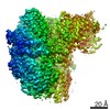
X-RAY DIFFRACTION / SYNCHROTRON / MAD / Resolution: 2.9 Å
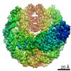
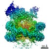
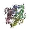
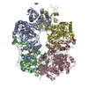
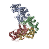
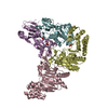
A: ATP phosphoribosyltransferase regulatory subunit B: ATP phosphoribosyltransferase regulatory subunit C: ATP phosphoribosyltransferase regulatory subunit D: ATP phosphoribosyltransferase regulatory subunit E: ATP phosphoribosyltransferase F: ATP phosphoribosyltransferase G: ATP phosphoribosyltransferase H: ATP phosphoribosyltransferase hetero molecules
A: ATP phosphoribosyltransferase regulatory subunit B: ATP phosphoribosyltransferase regulatory subunit G: ATP phosphoribosyltransferase H: ATP phosphoribosyltransferase hetero molecules
C: ATP phosphoribosyltransferase regulatory subunit D: ATP phosphoribosyltransferase regulatory subunit E: ATP phosphoribosyltransferase F: ATP phosphoribosyltransferase hetero molecules
In the structure databanks used in Yorodumi, some data are registered as the other names, "COVID-19 virus" and "2019-nCoV". Here are the details of the virus and the list of structure data.
Jan 31, 2019. EMDB accession codes are about to change! (news from PDBe EMDB page)
EMDB accession codes are about to change! (news from PDBe EMDB page)
The allocation of 4 digits for EMDB accession codes will soon come to an end. Whilst these codes will remain in use, new EMDB accession codes will include an additional digit and will expand incrementally as the available range of codes is exhausted. The current 4-digit format prefixed with “EMD-” (i.e. EMD-XXXX) will advance to a 5-digit format (i.e. EMD-XXXXX), and so on. It is currently estimated that the 4-digit codes will be depleted around Spring 2019, at which point the 5-digit format will come into force.
The EM Navigator/Yorodumi systems omit the EMD- prefix.
Related info.:Q: What is EMD? / ID/Accession-code notation in Yorodumi/EM Navigator
Yorodumi is a browser for structure data from EMDB, PDB, SASBDB, etc.
This page is also the successor to EM Navigator detail page, and also detail information page/front-end page for Omokage search.
The word "yorodu" (or yorozu) is an old Japanese word meaning "ten thousand". "mi" (miru) is to see.
Related info.:EMDB / PDB / SASBDB / Comparison of 3 databanks / Yorodumi Search / Aug 31, 2016. New EM Navigator & Yorodumi / Yorodumi Papers / Jmol/JSmol / Function and homology information / Changes in new EM Navigator and Yorodumi
 Movie
Movie Controller

 Yorodumi
 Open data
 Basic information
 Components
 Keywords
 Function and homology information
 Lactococcus lactis (lactic acid bacteria)
 X-RAY DIFFRACTION /
  Authors
 Citation
 Structure visualization
 Downloads & links
 Other downloads

 PDBj
PDBj
 Assembly
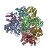

 Mass: 247.838 Da / Num. of mol.: 2 / Source method: obtained synthetically / Formula: WO4
 Mass: 94.971 Da / Num. of mol.: 6 / Source method: obtained synthetically / Formula: PO4
 Sample preparation
 Processing