 Movie
Movie Controller

[English] 日本語
 Yorodumi
- PDB-1z6k: Citrate lyase beta subunit complexed with oxaloacetate and magnes... -
+ Open data

- Basic information
| Entry | Database: PDB / ID: 1z6k | ||||||
|---|---|---|---|---|---|---|---|
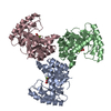
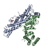
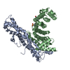
| Title | Citrate lyase beta subunit complexed with oxaloacetate and magnesium from M. tuberculosis | ||||||
 Components Components | Citrate Lyase beta subunit | ||||||
 Keywords Keywords | LYASE / TIM barrel / Structural Genomics / PSI / Protein Structure Initiative / TB Structural Genomics Consortium / TBSGC | ||||||
| Function / homology |  Function and homology information Function and homology informationLyases; Carbon-carbon lyases / oxaloacetate metabolic process / lyase activity / magnesium ion binding / metal ion binding Similarity search - Function | ||||||
| Biological species |   Mycobacterium tuberculosis (bacteria) Mycobacterium tuberculosis (bacteria) | ||||||
| Method |  X-RAY DIFFRACTION / SYNCHROTRON / MOLECULAR REPLACEMENT / Resolution: 2.3 Å X-RAY DIFFRACTION / SYNCHROTRON / MOLECULAR REPLACEMENT / Resolution: 2.3 Å | ||||||
 Authors Authors | Goulding, C.W. / Bowers, P.M. / Segelke, B. / Lekin, T. / Kim, C.Y. / Terwilliger, T.C. / Eisenberg, D. / TB Structural Genomics Consortium (TBSGC) | ||||||
 Citation Citation | Journal: J.Mol.Biol. / Year: 2007 Title: The structure and computational analysis of Mycobacterium tuberculosis protein CitE suggest a novel enzymatic function. Authors: Goulding, C.W. / Bowers, P.M. / Segelke, B. / Lekin, T. / Kim, C.Y. / Terwilliger, T.C. / Eisenberg, D. | ||||||
| History |
|
- Structure visualization
| Structure viewer | Molecule: MolmilJmol/JSmol |
|---|
- Downloads & links
-Download
| PDBx/mmCIF format | 1z6k.cif.gz | 57.7 KB | Display | PDBx/mmCIF format |
|---|---|---|---|---|
| PDB format | pdb1z6k.ent.gz | 40.5 KB | Display | PDB format |
| PDBx/mmJSON format | 1z6k.json.gz | Tree view | PDBx/mmJSON format | |
| Others |  Other downloads Other downloads |
-Validation report
| Arichive directory | https://data.pdbj.org/pub/pdb/validation_reports/z6/1z6kftp://data.pdbj.org/pub/pdb/validation_reports/z6/1z6k | HTTPS FTP |
|---|
-Related structure data
| Related structure data |  1u5hSC S: Starting model for refinement C: citing same article ( |
|---|---|
| Similar structure data | |
| Other databases |


-Links
 PDBj
PDBj


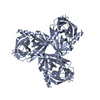
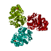
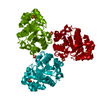
- Assembly
| Deposited unit | 
| |||||||||
|---|---|---|---|---|---|---|---|---|---|---|
| 1 | 
| |||||||||
| Unit cell |
| |||||||||
| Components on special symmetry positions |
|
-Components
| #1: Protein | Mass: 31420.410 Da / Num. of mol.: 1 Source method: isolated from a genetically manipulated source Source: (gene. exp.) Mycobacterium tuberculosis (bacteria) / Strain: H37Rv / Gene: Rv2498c / Plasmid: pET28d+ / Production host: References: UniProt: O06162, UniProt: P9WPE1*PLUS, citrate (pro-3S)-lyase |
|---|---|
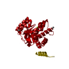
| #2: Chemical | ChemComp-OAA /   Mass: 131.064 Da / Num. of mol.: 1 / Source method: obtained synthetically / Formula: C4H3O5 Mass: 131.064 Da / Num. of mol.: 1 / Source method: obtained synthetically / Formula: C4H3O5 |
| #3: Chemical | ChemComp-MG /   Mass: 24.305 Da / Num. of mol.: 1 / Source method: obtained synthetically / Formula: Mg Mass: 24.305 Da / Num. of mol.: 1 / Source method: obtained synthetically / Formula: Mg |
| #4: Water | ChemComp-HOH /  Mass: 18.015 Da / Num. of mol.: 71 / Source method: isolated from a natural source / Formula: H2O Mass: 18.015 Da / Num. of mol.: 71 / Source method: isolated from a natural source / Formula: H2O |
| Has protein modification | Y |
-Experimental details
-Experiment
| Experiment | Method: X-RAY DIFFRACTION / Number of used crystals: 1 |
|---|
- Sample preparation
| Crystal | Density Matthews: 3.01 Å3/Da / Density % sol: 58.8 % |
|---|---|
| Crystal grow | Temperature: 300 K / Method: vapor diffusion, hanging drop / pH: 8.4 Details: sodium formate, DMSO., pH 8.4, VAPOR DIFFUSION, HANGING DROP, temperature 300K |
-Data collection
| Diffraction | Mean temperature: 200 K |
|---|---|
| Diffraction source | Source: SYNCHROTRON / Site: ALS  / Beamline: 5.0.2 / Wavelength: 1.5418 / Wavelength: 1.5418 Å / Beamline: 5.0.2 / Wavelength: 1.5418 / Wavelength: 1.5418 Å |
| Detector | Type: ADSC QUANTUM 4 / Detector: CCD / Date: Nov 5, 2004 |
| Radiation | Monochromator: SAGITALLY FOCUSED Si(111) / Protocol: SINGLE WAVELENGTH / Monochromatic (M) / Laue (L): M / Scattering type: x-ray |
| Radiation wavelength | Wavelength: 1.5418 Å / Relative weight: 1 |
| Reflection | Resolution: 2.3→100 Å / Num. all: 14893 / Num. obs: 14893 / % possible obs: 93 % / Observed criterion σ(F): 1 / Observed criterion σ(I): 1 |
- Processing
| Software |
| |||||||||||||||||||||||||
|---|---|---|---|---|---|---|---|---|---|---|---|---|---|---|---|---|---|---|---|---|---|---|---|---|---|---|
| Refinement | Method to determine structure: MOLECULAR REPLACEMENT Starting model: 1U5H Resolution: 2.3→10 Å / Isotropic thermal model: isotropic / σ(F): 2 / Stereochemistry target values: Engh & Huber
| |||||||||||||||||||||||||
| Refinement step | Cycle: LAST / Resolution: 2.3→10 Å
|
