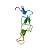
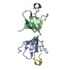
- PDB-1z6c: Solution structure of an EGF pair (EGF34) from vitamin K-dependen... -
+
データを開く
IDまたはキーワード:
読み込み中...
-
基本情報
登録情報
データベース: PDB / ID: 1z6c
タイトル
Solution structure of an EGF pair (EGF34) from vitamin K-dependent protein S
要素
Vitamin K-dependent protein S
キーワード
BLOOD CLOTTING / EGF module
機能・相同性
機能・相同性情報
endopeptidase inhibitor activity / Transport of gamma-carboxylated protein precursors from the endoplasmic reticulum to the Golgi apparatus / Gamma-carboxylation of protein precursors / Common Pathway of Fibrin Clot Formation / Removal of aminoterminal propeptides from gamma-carboxylated proteins / fibrinolysis / Intrinsic Pathway of Fibrin Clot Formation / platelet alpha granule lumen / Regulation of Complement cascade / Cell surface interactions at the vascular wall ...endopeptidase inhibitor activity / Transport of gamma-carboxylated protein precursors from the endoplasmic reticulum to the Golgi apparatus / Gamma-carboxylation of protein precursors / Common Pathway of Fibrin Clot Formation / Removal of aminoterminal propeptides from gamma-carboxylated proteins / fibrinolysis / Intrinsic Pathway of Fibrin Clot Formation / platelet alpha granule lumen / Regulation of Complement cascade / Cell surface interactions at the vascular wall / Golgi lumen / blood coagulation / Platelet degranulation / blood microparticle / positive regulation of phosphatidylinositol 3-kinase/protein kinase B signal transduction / Golgi membrane / calcium ion binding / endoplasmic reticulum membrane / extracellular space / extracellular exosome / extracellular region / plasma membrane 類似検索 - 分子機能
手法: Simulated annealing Torsion angle dynamics / ソフトェア番号: 1 詳細: The structures are based on a total of 912 restraints, 756 NOE restraints, 76 torsion angle restraints, 46 residual dipolar couplings and 30 restraints from hydrogen bonds
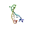
代表構造
選択基準: lowest energy
NMRアンサンブル
コンフォーマー選択の基準: structures with the lowest energy 計算したコンフォーマーの数: 200 / 登録したコンフォーマーの数: 20
 ムービー
ムービー コントローラー


 データを開く
 基本情報
基本情報 要素
要素 キーワード
キーワード 機能・相同性情報
 Homo sapiens (ヒト)
 データ登録者
 引用
引用 構造の表示
構造の表示 ダウンロードとリンク
 その他のダウンロード
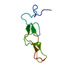


 PDBj
PDBj


 集合体
集合体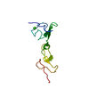


 分子量: 40.078 Da / 分子数: 2 / 由来タイプ: 合成 / 式: Ca
 分子量: 18.015 Da / 分子数: 2 / 由来タイプ: 天然 / 式: H2O
 試料調製
試料調製 解析
解析