- PDB-1z6c: Solution structure of an EGF pair (EGF34) from vitamin K-dependen... -
+
Open data
ID or keywords:
Loading...
-
Basic information
Entry
Database: PDB / ID: 1z6c
Title
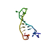
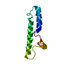
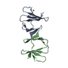
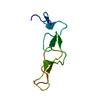
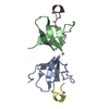
Solution structure of an EGF pair (EGF34) from vitamin K-dependent protein S
Components
Vitamin K-dependent protein S
Keywords
BLOOD CLOTTING / EGF module
Function / homology
Function and homology information
Dengue Virus Attachment and Entry / negative regulation of coagulation / endopeptidase inhibitor activity / Transport of gamma-carboxylated protein precursors from the endoplasmic reticulum to the Golgi apparatus / : / Gamma-carboxylation of protein precursors / Removal of aminoterminal propeptides from gamma-carboxylated proteins / fibrinolysis / : / platelet alpha granule lumen ...Dengue Virus Attachment and Entry / negative regulation of coagulation / endopeptidase inhibitor activity / Transport of gamma-carboxylated protein precursors from the endoplasmic reticulum to the Golgi apparatus / : / Gamma-carboxylation of protein precursors / Removal of aminoterminal propeptides from gamma-carboxylated proteins / fibrinolysis / : / platelet alpha granule lumen / Regulation of Complement cascade / Cell surface interactions at the vascular wall / Golgi lumen / blood coagulation / Platelet degranulation / blood microparticle / positive regulation of phosphatidylinositol 3-kinase/protein kinase B signal transduction / Golgi membrane / calcium ion binding / endoplasmic reticulum membrane / : / extracellular exosome / extracellular region / plasma membrane Similarity search - Function
Method: Simulated annealing Torsion angle dynamics / Software ordinal: 1 Details: The structures are based on a total of 912 restraints, 756 NOE restraints, 76 torsion angle restraints, 46 residual dipolar couplings and 30 restraints from hydrogen bonds
NMR representative
Selection criteria: lowest energy
NMR ensemble
Conformer selection criteria: structures with the lowest energy Conformers calculated total number: 200 / Conformers submitted total number: 20
+
About Yorodumi
-
News
-
Feb 9, 2022. New format data for meta-information of EMDB entries
New format data for meta-information of EMDB entries
Version 3 of the EMDB header file is now the official format.
The previous official version 1.9 will be removed from the archive.
In the structure databanks used in Yorodumi, some data are registered as the other names, "COVID-19 virus" and "2019-nCoV". Here are the details of the virus and the list of structure data.
Jan 31, 2019. EMDB accession codes are about to change! (news from PDBe EMDB page)
EMDB accession codes are about to change! (news from PDBe EMDB page)
The allocation of 4 digits for EMDB accession codes will soon come to an end. Whilst these codes will remain in use, new EMDB accession codes will include an additional digit and will expand incrementally as the available range of codes is exhausted. The current 4-digit format prefixed with “EMD-” (i.e. EMD-XXXX) will advance to a 5-digit format (i.e. EMD-XXXXX), and so on. It is currently estimated that the 4-digit codes will be depleted around Spring 2019, at which point the 5-digit format will come into force.
The EM Navigator/Yorodumi systems omit the EMD- prefix.
Related info.:Q: What is EMD? / ID/Accession-code notation in Yorodumi/EM Navigator
Yorodumi is a browser for structure data from EMDB, PDB, SASBDB, etc.
This page is also the successor to EM Navigator detail page, and also detail information page/front-end page for Omokage search.
The word "yorodu" (or yorozu) is an old Japanese word meaning "ten thousand". "mi" (miru) is to see.
Related info.:EMDB / PDB / SASBDB / Comparison of 3 databanks / Yorodumi Search / Aug 31, 2016. New EM Navigator & Yorodumi / Yorodumi Papers / Jmol/JSmol / Function and homology information / Changes in new EM Navigator and Yorodumi
 Movie
Movie Controller

 Yorodumi
 Open data
 Basic information
 Components
 Keywords
 Function and homology information
 Homo sapiens (human)
 Authors
 Citation
 Structure visualization
 Downloads & links
 Other downloads
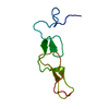


 PDBj
PDBj


 Assembly
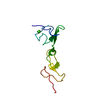


 Mass: 40.078 Da / Num. of mol.: 2 / Source method: obtained synthetically / Formula: Ca
 Mass: 18.015 Da / Num. of mol.: 2 / Source method: isolated from a natural source / Formula: H2O
 Sample preparation
 Processing