 Movie
Movie Controller

[English] 日本語
 Yorodumi
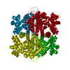
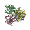
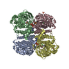
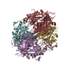
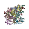
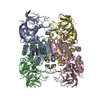
- PDB-1z69: Crystal structure of methylenetetrahydromethanopterin reductase (... -
+ Open data

- Basic information
| Entry | Database: PDB / ID: 1z69 | ||||||
|---|---|---|---|---|---|---|---|
| Title | Crystal structure of methylenetetrahydromethanopterin reductase (Mer) in complex with coenzyme F420 | ||||||
 Components Components | Coenzyme F420-dependent N(5),N(10)-methylenetetrahydromethanopterin reductase | ||||||
 Keywords Keywords | OXIDOREDUCTASE / (alpha / beta)8 barrel | ||||||
| Function / homology |  Function and homology information Function and homology information5,10-methylenetetrahydromethanopterin reductase / coenzyme F420-dependent N5,N10-methenyltetrahydromethanopterin reductase activity / methanogenesis, from carbon dioxide / oxidoreductase activity, acting on paired donors, with incorporation or reduction of molecular oxygen / one-carbon metabolic process / cytoplasm Similarity search - Function | ||||||
| Biological species |  Methanosarcina barkeri (archaea) Methanosarcina barkeri (archaea) | ||||||
| Method |  X-RAY DIFFRACTION / SYNCHROTRON / MOLECULAR REPLACEMENT / Resolution: 2.61 Å X-RAY DIFFRACTION / SYNCHROTRON / MOLECULAR REPLACEMENT / Resolution: 2.61 Å | ||||||
 Authors Authors | Aufhammer, S.W. / Warkentin, E. / Ermler, U. / Hagemeier, C.H. / Thauer, R.K. / Shima, S. | ||||||
 Citation Citation | Journal: Protein Sci. / Year: 2005 Title: Crystal structure of methylenetetrahydromethanopterin reductase (Mer) in complex with coenzyme F420: Architecture of the F420/FMN binding site of enzymes within the nonprolyl cis-peptide ...Title: Crystal structure of methylenetetrahydromethanopterin reductase (Mer) in complex with coenzyme F420: Architecture of the F420/FMN binding site of enzymes within the nonprolyl cis-peptide containing bacterial luciferase family Authors: Aufhammer, S.W. / Warkentin, E. / Ermler, U. / Hagemeier, C.H. / Thauer, R.K. / Shima, S. | ||||||
| History |
|
- Structure visualization
| Structure viewer | Molecule: MolmilJmol/JSmol |
|---|
- Downloads & links
-Download
| PDBx/mmCIF format | 1z69.cif.gz | 256.5 KB | Display | PDBx/mmCIF format |
|---|---|---|---|---|
| PDB format | pdb1z69.ent.gz | 206.5 KB | Display | PDB format |
| PDBx/mmJSON format | 1z69.json.gz | Tree view | PDBx/mmJSON format | |
| Others |  Other downloads Other downloads |
-Validation report
| Arichive directory | https://data.pdbj.org/pub/pdb/validation_reports/z6/1z69ftp://data.pdbj.org/pub/pdb/validation_reports/z6/1z69 | HTTPS FTP |
|---|
-Related structure data
| Related structure data | 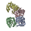 1f07S S: Starting model for refinement |
|---|---|
| Similar structure data |
-Links
 PDBj
PDBj
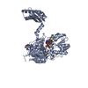
- Assembly
| Deposited unit | 
| ||||||||||
|---|---|---|---|---|---|---|---|---|---|---|---|
| 1 |
| ||||||||||
| Unit cell |
|
-Components
| #1: Protein | Mass: 34501.938 Da / Num. of mol.: 4 / Source method: isolated from a natural source / Source: (natural) Methanosarcina barkeri (archaea) / Strain: Fusaro (DSMZ 804)References: GenBank: 48840204, UniProt: Q46FV4*PLUS, EC: 1.5.99.11 #2: Chemical | ChemComp-F42 / 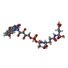  Mass: 773.593 Da / Num. of mol.: 4 / Source method: obtained synthetically / Formula: C29H36N5O18P Mass: 773.593 Da / Num. of mol.: 4 / Source method: obtained synthetically / Formula: C29H36N5O18P#3: Chemical |   Mass: 35.453 Da / Num. of mol.: 2 / Source method: obtained synthetically / Formula: Cl Mass: 35.453 Da / Num. of mol.: 2 / Source method: obtained synthetically / Formula: Cl#4: Chemical | ChemComp-1PG / | 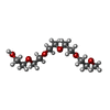  Mass: 252.305 Da / Num. of mol.: 1 / Source method: obtained synthetically / Formula: C11H24O6 Mass: 252.305 Da / Num. of mol.: 1 / Source method: obtained synthetically / Formula: C11H24O6#5: Water | ChemComp-HOH / |  Mass: 18.015 Da / Num. of mol.: 239 / Source method: isolated from a natural source / Formula: H2O Mass: 18.015 Da / Num. of mol.: 239 / Source method: isolated from a natural source / Formula: H2O |
|---|
-Experimental details
-Experiment
| Experiment | Method: X-RAY DIFFRACTION / Number of used crystals: 1 |
|---|
- Sample preparation
| Crystal | Density Matthews: 2.38 Å3/Da / Density % sol: 46.7 % |
|---|---|
| Crystal grow | Temperature: 281 K / Method: vapor diffusion, hanging drop / pH: 8.5 Details: Tris/HCl, PEG 4000, Isopropanol, pH 8.5, VAPOR DIFFUSION, HANGING DROP, temperature 281K |
-Data collection
| Diffraction | Mean temperature: 100 K |
|---|---|
| Diffraction source | Source: SYNCHROTRON / Site: ESRF  / Beamline: ID29 / Wavelength: 0.9792 Å / Beamline: ID29 / Wavelength: 0.9792 Å |
| Detector | Type: ADSC QUANTUM 4 / Detector: CCD / Date: Feb 9, 2004 |
| Radiation | Monochromator: Si(111) / Protocol: SINGLE WAVELENGTH / Monochromatic (M) / Laue (L): M / Scattering type: x-ray |
| Radiation wavelength | Wavelength: 0.9792 Å / Relative weight: 1 |
| Reflection | Resolution: 2.6→30 Å / Num. all: 40607 / Num. obs: 40607 / % possible obs: 97.1 % / Observed criterion σ(F): 0 / Observed criterion σ(I): 0 / Redundancy: 3.96 % / Biso Wilson estimate: 37.7 Å2 / Rmerge(I) obs: 0.085 / Net I/σ(I): 14.2 |
| Reflection shell | Resolution: 2.6→2.74 Å / Redundancy: 3.6 % / Rmerge(I) obs: 0.214 / Mean I/σ(I) obs: 6.8 / Num. unique all: 5785 / % possible all: 86.4 |
- Processing
| Software |
| ||||||||||||||||||||||||||||||||||||
|---|---|---|---|---|---|---|---|---|---|---|---|---|---|---|---|---|---|---|---|---|---|---|---|---|---|---|---|---|---|---|---|---|---|---|---|---|---|
| Refinement | Method to determine structure: MOLECULAR REPLACEMENT Starting model: PDB entry 1F07 Resolution: 2.61→19.95 Å / Rfactor Rfree error: 0.005 / Data cutoff high absF: 208649.04 / Data cutoff low absF: 0 / Isotropic thermal model: RESTRAINED / Cross valid method: THROUGHOUT / σ(F): 0 / Stereochemistry target values: Engh & Huber Details: SEE REMARK 11 NCS RESTRAINTS NCS RESTRAINTS WERE USED FOR ALL FOUR CHAINS EXCEPT FOR RESIDUES 214 - 283 OF CHAIN D. DIFFERENT WEIGHTS WERE APPLIED TO MAIN CHAIN AND SIDE CHAINS. MAIN CHAIN ...Details: SEE REMARK 11 NCS RESTRAINTS NCS RESTRAINTS WERE USED FOR ALL FOUR CHAINS EXCEPT FOR RESIDUES 214 - 283 OF CHAIN D. DIFFERENT WEIGHTS WERE APPLIED TO MAIN CHAIN AND SIDE CHAINS. MAIN CHAIN ATOMS, EXCEPT FOR RESIDUES 214-285 CHAIN A CHAIN B CHAIN D RMSD 0.058 0.057 0.066 (A) REFERENCE MC ATOMS OF CHAIN C SIDE CHAIN ATOMS, EXCEPT FOR RESIDUES 214-285 CHAIN A CHAIN B CHAIN D RMSD 0.12 0.12 0.13 (A) REFERENCE SC ATOMS OF CHAIN C MAIN CHAIN ATOMS, RESIDUES 214-285 CHAIN A CHAIN B CHAIN D RMSD 0.056 0.054 --- (A) REFERENCE MC ATOMS OF CHAIN C SIDE CHAIN ATOMS, RESIDUES 214-285 CHAIN A CHAIN B CHAIN D RMSD 0.14 0.15 --- (A) REFERENCE SC ATOMS OF CHAIN C
| ||||||||||||||||||||||||||||||||||||
| Solvent computation | Solvent model: FLAT MODEL / Bsol: 37.7686 Å2 / ksol: 0.36323 e/Å3 | ||||||||||||||||||||||||||||||||||||
| Displacement parameters | Biso mean: 28.7 Å2
| ||||||||||||||||||||||||||||||||||||
| Refine analyze |
| ||||||||||||||||||||||||||||||||||||
| Refinement step | Cycle: LAST / Resolution: 2.61→19.95 Å
| ||||||||||||||||||||||||||||||||||||
| Refine LS restraints |
| ||||||||||||||||||||||||||||||||||||
| LS refinement shell | Resolution: 2.61→2.76 Å / Rfactor Rfree error: 0.018 / Total num. of bins used: 6
| ||||||||||||||||||||||||||||||||||||
| Xplor file |
|
