 Movie
Movie Controller

[English] 日本語
 Yorodumi
- PDB-1z5x: hemipteran ecdysone receptor ligand-binding domain complexed with... -
+ Open data

- Basic information
| Entry | Database: PDB / ID: 1z5x | ||||||
|---|---|---|---|---|---|---|---|
| Title | hemipteran ecdysone receptor ligand-binding domain complexed with ponasterone A | ||||||
 Components Components |
| ||||||
 Keywords Keywords | Hormone/Growth Factor Receptor / ecdysone receptor / ponasterone A / nuclear receptor / ECR / USP / ecdysone / Hormone-Growth Factor Receptor COMPLEX | ||||||
| Function / homology | Retinoid X Receptor / Retinoid X Receptor / Orthogonal Bundle / Mainly Alpha / 2,3,14,20,22-PENTAHYDROXYCHOLEST-7-EN-6-ONE / PHOSPHATE ION Function and homology information Function and homology information | ||||||
| Biological species |  Bemisia tabaci (sweet potato whitefly) Bemisia tabaci (sweet potato whitefly) | ||||||
| Method |  X-RAY DIFFRACTION / MOLECULAR REPLACEMENT / Resolution: 3.07 Å X-RAY DIFFRACTION / MOLECULAR REPLACEMENT / Resolution: 3.07 Å | ||||||
 Authors Authors | Carmichael, J.A. / Lawrence, M.C. / Graham, L.D. / Pilling, P.A. / Epa, V.C. / Noyce, L. / Lovrecz, G. / Winkler, D.A. / Pawlak-Skrzecz, A. | ||||||
 Citation Citation | Journal: J.Biol.Chem. / Year: 2005 Title: The X-ray structure of a hemipteran ecdysone receptor ligand-binding domain: comparison with a lepidopteran ecdysone receptor ligand-binding domain and implications for insecticide design. Authors: Carmichael, J.A. / Lawrence, M.C. / Graham, L.D. / Pilling, P.A. / Epa, V.C. / Noyce, L. / Lovrecz, G. / Winkler, D.A. / Pawlak-Skrzecz, A. / Eaton, R.E. / Hannan, G.N. / Hill, R.J. | ||||||
| History |
| ||||||
| Remark 999 | SEQUENCE According to authors the N-termini of the crystal contents could not be verified. SDS-PAGE ...SEQUENCE According to authors the N-termini of the crystal contents could not be verified. SDS-PAGE analysis of crystals and stored protein revealed polypeptides of lower molecular weight than expected, presumably the result of progressive proteolysis. N-terminal sequencing of major bands extracted from SDS-PAGE could only confirm one band as corresponding to a sequence starting at residue LEU U 285. At the time of processing there were no suitable database references for chains U or E |
- Structure visualization
| Structure viewer | Molecule: MolmilJmol/JSmol |
|---|
- Downloads & links
-Download
| PDBx/mmCIF format | 1z5x.cif.gz | 105 KB | Display | PDBx/mmCIF format |
|---|---|---|---|---|
| PDB format | pdb1z5x.ent.gz | 78.2 KB | Display | PDB format |
| PDBx/mmJSON format | 1z5x.json.gz | Tree view | PDBx/mmJSON format | |
| Others |  Other downloads Other downloads |
-Validation report
| Arichive directory | https://data.pdbj.org/pub/pdb/validation_reports/z5/1z5xftp://data.pdbj.org/pub/pdb/validation_reports/z5/1z5x | HTTPS FTP |
|---|
-Related structure data
| Related structure data | 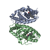 1dkfS S: Starting model for refinement |
|---|---|
| Similar structure data |


-Links
 PDBj
PDBj- Assembly
| Deposited unit | 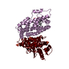
| ||||||||
|---|---|---|---|---|---|---|---|---|---|
| 1 |
| ||||||||
| Unit cell |
| ||||||||
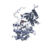
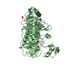
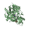
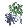
| Details | the biological assembly is a heterodimer of Usp (chain U) and Ecr (chain E) |
-Components
| #1: Protein | Mass: 29997.205 Da / Num. of mol.: 1 Source method: isolated from a genetically manipulated source Source: (gene. exp.) Bemisia tabaci (sweet potato whitefly) / Gene: Usp / Production host: Trichoplusia ni (cabbage looper) / Strain (production host): Hi-5 (Invitrogen) | ||
|---|---|---|---|
| #2: Protein | Mass: 35873.070 Da / Num. of mol.: 1 / Fragment: ligand binding subunit Source method: isolated from a genetically manipulated source Source: (gene. exp.) Bemisia tabaci (sweet potato whitefly) / Gene: Ecr / Production host: Trichoplusia ni (cabbage looper) / Strain (production host): Hi-5 (Invitrogen) | ||
| #3: Chemical | ChemComp-PO4 /   Mass: 94.971 Da / Num. of mol.: 4 / Source method: obtained synthetically / Formula: PO4 Mass: 94.971 Da / Num. of mol.: 4 / Source method: obtained synthetically / Formula: PO4#4: Chemical | ChemComp-P1A / | 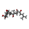  Mass: 464.635 Da / Num. of mol.: 1 / Source method: obtained synthetically / Formula: C27H44O6 Mass: 464.635 Da / Num. of mol.: 1 / Source method: obtained synthetically / Formula: C27H44O6 |
-Experimental details
-Experiment
| Experiment | Method: X-RAY DIFFRACTION / Number of used crystals: 1 |
|---|
- Sample preparation
| Crystal | Density Matthews: 4.35 Å3/Da / Density % sol: 71.5 % |
|---|---|
| Crystal grow | Temperature: 298 K / Method: vapor diffusion, hanging drop / pH: 7.5 Details: 1 M ammonium phosphate, 4.5 % trehalose, 10 mM DTT, 10 % glycerol, 3 mM ponasterone A, 0.1 M HEPES, pH 7.5, VAPOR DIFFUSION, HANGING DROP, temperature 298K |
-Data collection
| Diffraction | Mean temperature: 115 K |
|---|---|
| Diffraction source | Source: ROTATING ANODE / Type: RIGAKU / Wavelength: 1.5418 Å |
| Detector | Type: RIGAKU RAXIS IV / Detector: IMAGE PLATE / Date: Feb 21, 2002 / Details: Yale-MSC mirrors |
| Radiation | Protocol: SINGLE WAVELENGTH / Monochromatic (M) / Laue (L): M / Scattering type: x-ray |
| Radiation wavelength | Wavelength: 1.5418 Å / Relative weight: 1 |
| Reflection | Resolution: 3.07→27.48 Å / Num. all: 16756 / Num. obs: 16756 / % possible obs: 99.7 % / Observed criterion σ(F): 0 / Observed criterion σ(I): 0 / Redundancy: 11.4 % / Rsym value: 0.13 / Net I/σ(I): 19.3 |
| Reflection shell | Resolution: 3.07→3.11 Å / Redundancy: 10.5 % / Mean I/σ(I) obs: 5.6 / Num. unique all: 2713 / Rsym value: 0.51 / % possible all: 99 |
- Processing
| Software |
| ||||||||||||||||||||||||||||||||||||||||||||||||||||||||||||
|---|---|---|---|---|---|---|---|---|---|---|---|---|---|---|---|---|---|---|---|---|---|---|---|---|---|---|---|---|---|---|---|---|---|---|---|---|---|---|---|---|---|---|---|---|---|---|---|---|---|---|---|---|---|---|---|---|---|---|---|---|---|
| Refinement | Method to determine structure: MOLECULAR REPLACEMENT Starting model: Homology model derived from pdb entry 1DKF Resolution: 3.07→27.48 Å / Rfactor Rfree error: 0.009 / Data cutoff high absF: 2256128.06 / Data cutoff low absF: 0 / Isotropic thermal model: RESTRAINED / Cross valid method: THROUGHOUT / σ(F): 0 / σ(I): 0 / Stereochemistry target values: Engh & Huber
| ||||||||||||||||||||||||||||||||||||||||||||||||||||||||||||
| Solvent computation | Solvent model: FLAT MODEL / Bsol: 24.4674 Å2 / ksol: 0.364873 e/Å3 | ||||||||||||||||||||||||||||||||||||||||||||||||||||||||||||
| Displacement parameters | Biso mean: 46.2 Å2
| ||||||||||||||||||||||||||||||||||||||||||||||||||||||||||||
| Refine analyze |
| ||||||||||||||||||||||||||||||||||||||||||||||||||||||||||||
| Refinement step | Cycle: LAST / Resolution: 3.07→27.48 Å
| ||||||||||||||||||||||||||||||||||||||||||||||||||||||||||||
| Refine LS restraints |
| ||||||||||||||||||||||||||||||||||||||||||||||||||||||||||||
| LS refinement shell | Resolution: 3.07→3.26 Å / Rfactor Rfree error: 0.029 / Total num. of bins used: 6
| ||||||||||||||||||||||||||||||||||||||||||||||||||||||||||||
| Xplor file |
|
