 Movie
Movie Controller

[English] 日本語
 Yorodumi
- PDB-1z5u: Crystal structure of S. typhimurium AphA complexed with cyclic-AMP -
+ Open data

- Basic information
| Entry | Database: PDB / ID: 1z5u | ||||||
|---|---|---|---|---|---|---|---|
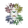
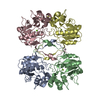
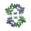
| Title | Crystal structure of S. typhimurium AphA complexed with cyclic-AMP | ||||||
 Components Components | AphA protein | ||||||
 Keywords Keywords | HYDROLASE / Class-B bacterial acid phosphatase / cyclic-AMP complex of AphA protein / metalloenzyme | ||||||
| Function / homology |  Function and homology information Function and homology informationacid phosphatase / acid phosphatase activity / outer membrane-bounded periplasmic space / metal ion binding Similarity search - Function | ||||||
| Biological species |  Salmonella typhimurium (bacteria) Salmonella typhimurium (bacteria) | ||||||
| Method |  X-RAY DIFFRACTION / FOURIER SYNTHESIS / Resolution: 2.3 Å X-RAY DIFFRACTION / FOURIER SYNTHESIS / Resolution: 2.3 Å | ||||||
 Authors Authors | Makde, R.D. / Kumar, V. | ||||||
 Citation Citation | Journal: To be Published Title: Structure and Function of class-B bacterial non-specific acid phosphatase: functional role of invariant Lys154 Authors: Makde, R.D. / Gupta, G.D. / Kumar, V. #1: Journal: ACTA CRYSTALLOGR.,SECT.D / Year: 2003 Title: Expression, purification, crystallization and preliminary X-ray diffraction studies of recombinant class B non-specific acid phosphatase of Salmonella typhimurium Authors: Makde, R.D. / Kumar, V. / Gupta, G.D. / Jasti, J. / Singh, T.P. / Mahajan, S.K. | ||||||
| History |
|
- Structure visualization
| Structure viewer | Molecule: MolmilJmol/JSmol |
|---|
- Downloads & links
-Download
| PDBx/mmCIF format | 1z5u.cif.gz | 187 KB | Display | PDBx/mmCIF format |
|---|---|---|---|---|
| PDB format | pdb1z5u.ent.gz | 148.1 KB | Display | PDB format |
| PDBx/mmJSON format | 1z5u.json.gz | Tree view | PDBx/mmJSON format | |
| Others |  Other downloads Other downloads |
-Validation report
| Arichive directory | https://data.pdbj.org/pub/pdb/validation_reports/z5/1z5uftp://data.pdbj.org/pub/pdb/validation_reports/z5/1z5u | HTTPS FTP |
|---|
-Related structure data
| Related structure data | 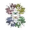 1z5gS S: Starting model for refinement |
|---|---|
| Similar structure data |

-Links
 PDBj
PDBj


- Assembly
| Deposited unit | 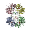
| ||||||||
|---|---|---|---|---|---|---|---|---|---|
| 1 |
| ||||||||
| Unit cell |
|
-Components
| #1: Protein | Mass: 23870.703 Da / Num. of mol.: 4 Source method: isolated from a genetically manipulated source Source: (gene. exp.) Salmonella typhimurium (bacteria) / Gene: aphA / Plasmid: pET21(a) / Species (production host): Escherichia coli / Production host: References: GenBank: 56694136, UniProt: Q540U1*PLUS, acid phosphatase #2: Chemical | ChemComp-MG /   Mass: 24.305 Da / Num. of mol.: 4 / Source method: obtained synthetically / Formula: Mg Mass: 24.305 Da / Num. of mol.: 4 / Source method: obtained synthetically / Formula: Mg#3: Chemical | ChemComp-CMP /   Mass: 329.206 Da / Num. of mol.: 4 / Source method: obtained synthetically / Formula: C10H12N5O6P Mass: 329.206 Da / Num. of mol.: 4 / Source method: obtained synthetically / Formula: C10H12N5O6P#4: Water | ChemComp-HOH / |  Mass: 18.015 Da / Num. of mol.: 476 / Source method: isolated from a natural source / Formula: H2O Mass: 18.015 Da / Num. of mol.: 476 / Source method: isolated from a natural source / Formula: H2O |
|---|
-Experimental details
-Experiment
| Experiment | Method: X-RAY DIFFRACTION / Number of used crystals: 1 |
|---|
- Sample preparation
| Crystal | Density Matthews: 2.33 Å3/Da / Density % sol: 47.2 % |
|---|---|
| Crystal grow | Temperature: 283 K / Method: vapor diffusion, sitting drop / pH: 4.7 Details: PEG6000, magnesium chloride, glycerol, sodium actetate, cyclic-AMP, pH 4.7, VAPOR DIFFUSION, SITTING DROP, temperature 283K |
-Data collection
| Diffraction | Mean temperature: 293 K |
|---|---|
| Diffraction source | Source: ROTATING ANODE / Type: RIGAKU RU200 / Wavelength: 1.5418 Å |
| Detector | Type: MARRESEARCH / Detector: IMAGE PLATE / Date: Mar 19, 2004 / Details: OSMIC MIRRORS |
| Radiation | Monochromator: graphite / Protocol: SINGLE WAVELENGTH / Monochromatic (M) / Laue (L): M / Scattering type: x-ray |
| Radiation wavelength | Wavelength: 1.5418 Å / Relative weight: 1 |
| Reflection | Resolution: 2.3→30 Å / Num. all: 38398 / Num. obs: 38398 / % possible obs: 94.8 % / Observed criterion σ(I): 0 / Redundancy: 3 % / Biso Wilson estimate: 32.5 Å2 / Rsym value: 0.081 / Net I/σ(I): 7.1 |
| Reflection shell | Resolution: 2.3→2.42 Å / % possible obs: 96.2 % / Redundancy: 2.9 % / Mean I/σ(I) obs: 2.7 / Num. unique all: 5619 / Rsym value: 0.269 / % possible all: 96.2 |
- Processing
| Software |
| |||||||||||||||||||||||||||
|---|---|---|---|---|---|---|---|---|---|---|---|---|---|---|---|---|---|---|---|---|---|---|---|---|---|---|---|---|
| Refinement | Method to determine structure: FOURIER SYNTHESIS Starting model: PDB CODE 1Z5G Resolution: 2.3→20 Å / Isotropic thermal model: ISOTROPIC / Cross valid method: THROUGHOUT / σ(F): 0 / Stereochemistry target values: Engh & Huber
| |||||||||||||||||||||||||||
| Solvent computation | Solvent model: Flat Model / Bsol: 32.18 Å2 / ksol: 0.311 e/Å3 | |||||||||||||||||||||||||||
| Displacement parameters | Biso mean: 29.7 Å2 | |||||||||||||||||||||||||||
| Refine analyze |
| |||||||||||||||||||||||||||
| Refinement step | Cycle: LAST / Resolution: 2.3→20 Å
| |||||||||||||||||||||||||||
| Refine LS restraints |
| |||||||||||||||||||||||||||
| LS refinement shell | Resolution: 2.3→2.38 Å / Total num. of bins used: 10
| |||||||||||||||||||||||||||
| Xplor file |
|
