 Movie
Movie Controller

[English] 日本語
 Yorodumi
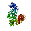
- PDB-1z5h: Crystal structures of the Tricorn interacting Factor F3 from Ther... -
+ Open data

- Basic information
| Entry | Database: PDB / ID: 1z5h | ||||||
|---|---|---|---|---|---|---|---|
| Title | Crystal structures of the Tricorn interacting Factor F3 from Thermoplasma acidophilum | ||||||
 Components Components | Tricorn protease interacting factor F3 | ||||||
 Keywords Keywords | HYDROLASE / Zinc aminopeptidase / Gluzicins / Tricorn protease / Superhelix | ||||||
| Function / homology |  Function and homology information Function and homology informationHydrolases; Acting on peptide bonds (peptidases); Aminopeptidases / peptide catabolic process / metalloaminopeptidase activity / peptide binding / proteolysis / : / zinc ion binding / membrane / cytoplasm Similarity search - Function | ||||||
| Biological species |   Thermoplasma acidophilum (acidophilic) Thermoplasma acidophilum (acidophilic) | ||||||
| Method |  X-RAY DIFFRACTION / SYNCHROTRON / MOLECULAR REPLACEMENT / Resolution: 2.3 Å X-RAY DIFFRACTION / SYNCHROTRON / MOLECULAR REPLACEMENT / Resolution: 2.3 Å | ||||||
 Authors Authors | Kyrieleis, O.J.P. / Goettig, P. / Kiefersauer, R. / Huber, R. / Brandstetter, H. | ||||||
 Citation Citation | Journal: J.MOL.BIOL. / Year: 2005 Title: Crystal Structures of the Tricorn Interacting Factor F3 from Thermoplasma acidophilum, a Zinc Aminopeptidase in Three Different Conformations Authors: Kyrieleis, O.J.P. / Goettig, P. / Kiefersauer, R. / Huber, R. / Brandstetter, H. | ||||||
| History |
|
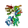
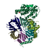
- Structure visualization
| Structure viewer | Molecule: MolmilJmol/JSmol |
|---|
- Downloads & links
-Download
| PDBx/mmCIF format | 1z5h.cif.gz | 336.2 KB | Display | PDBx/mmCIF format |
|---|---|---|---|---|
| PDB format | pdb1z5h.ent.gz | 271.4 KB | Display | PDB format |
| PDBx/mmJSON format | 1z5h.json.gz | Tree view | PDBx/mmJSON format | |
| Others |  Other downloads Other downloads |
-Validation report
| Arichive directory | https://data.pdbj.org/pub/pdb/validation_reports/z5/1z5hftp://data.pdbj.org/pub/pdb/validation_reports/z5/1z5h | HTTPS FTP |
|---|
-Related structure data


-Links
 PDBj
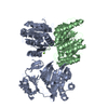
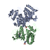
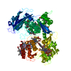
PDBj- Assembly
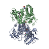
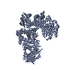
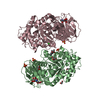
| Deposited unit | 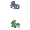
| ||||||||
|---|---|---|---|---|---|---|---|---|---|
| 1 | 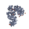
| ||||||||
| 2 | 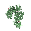
| ||||||||
| 3 | 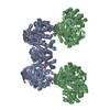
| ||||||||
| 4 | 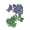
| ||||||||
| Unit cell |
|
-Components
| #1: Protein | Mass: 89492.727 Da / Num. of mol.: 2 Source method: isolated from a genetically manipulated source Source: (gene. exp.) Thermoplasma acidophilum (acidophilic) / Plasmid: pRset6c / Production host:  References: UniProt: O93655, Hydrolases; Acting on peptide bonds (peptidases); Aminopeptidases #2: Chemical | ChemComp-SO4 /   Mass: 96.063 Da / Num. of mol.: 17 / Source method: obtained synthetically / Formula: SO4 Mass: 96.063 Da / Num. of mol.: 17 / Source method: obtained synthetically / Formula: SO4#3: Chemical |   Mass: 65.409 Da / Num. of mol.: 2 / Source method: obtained synthetically / Formula: Zn Mass: 65.409 Da / Num. of mol.: 2 / Source method: obtained synthetically / Formula: Zn#4: Water | ChemComp-HOH / |  Mass: 18.015 Da / Num. of mol.: 779 / Source method: isolated from a natural source / Formula: H2O Mass: 18.015 Da / Num. of mol.: 779 / Source method: isolated from a natural source / Formula: H2O |
|---|
-Experimental details
-Experiment
| Experiment | Method: X-RAY DIFFRACTION / Number of used crystals: 1 |
|---|
- Sample preparation
| Crystal | Density Matthews: 3.11 Å3/Da / Density % sol: 60.51 % |
|---|---|
| Crystal grow | Temperature: 291 K / Method: vapor diffusion, sitting drop / pH: 8.5 Details: PEG 2000, lithium sulphate, TRIS, pH 8.5, VAPOR DIFFUSION, SITTING DROP, temperature 291K |
-Data collection
| Diffraction | Mean temperature: 100 K |
|---|---|
| Diffraction source | Source: SYNCHROTRON / Site: MPG/DESY, HAMBURG  / Beamline: BW6 / Wavelength: 1.005 Å / Beamline: BW6 / Wavelength: 1.005 Å |
| Detector | Type: MARRESEARCH / Detector: CCD / Date: Oct 16, 2003 |
| Radiation | Monochromator: graphit / Protocol: SINGLE WAVELENGTH / Monochromatic (M) / Laue (L): M / Scattering type: x-ray |
| Radiation wavelength | Wavelength: 1.005 Å / Relative weight: 1 |
| Reflection | Resolution: 2.3→19.5 Å / Num. all: 99537 / Num. obs: 99516 / % possible obs: 99.9 % / Biso Wilson estimate: 24.6 Å2 |
| Reflection shell | Resolution: 2.3→2.44 Å / % possible all: 100 |
- Processing
| Software |
| |||||||||||||||||||||||||
|---|---|---|---|---|---|---|---|---|---|---|---|---|---|---|---|---|---|---|---|---|---|---|---|---|---|---|
| Refinement | Method to determine structure: MOLECULAR REPLACEMENT / Resolution: 2.3→19.48 Å / Rfactor Rfree error: 0.004 / Data cutoff high absF: 4087184.01 / Data cutoff low absF: 0 / Isotropic thermal model: RESTRAINED / Cross valid method: THROUGHOUT / σ(F): 0 / Stereochemistry target values: Engh & Huber
| |||||||||||||||||||||||||
| Solvent computation | Solvent model: FLAT MODEL / Bsol: 37.8232 Å2 / ksol: 0.343331 e/Å3 | |||||||||||||||||||||||||
| Displacement parameters | Biso mean: 38.5 Å2
| |||||||||||||||||||||||||
| Refine analyze |
| |||||||||||||||||||||||||
| Refinement step | Cycle: LAST / Resolution: 2.3→19.48 Å
| |||||||||||||||||||||||||
| Refine LS restraints |
| |||||||||||||||||||||||||
| LS refinement shell | Resolution: 2.3→2.44 Å / Rfactor Rfree error: 0.012 / Total num. of bins used: 6
| |||||||||||||||||||||||||
| Xplor file |
|
