[English] 日本語
Yorodumi
- PDB-1z2m: Crystal Structure of ISG15, the Interferon-Induced Ubiquitin Cros... -

+
Open data


ID or keywords:

Loading...

-
Basic information

Entry
Database: PDB / ID: 1z2m
Title
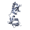
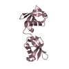
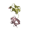
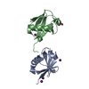
Crystal Structure of ISG15, the Interferon-Induced Ubiquitin Cross Reactive Protein
Componentsinterferon, alpha-inducible protein (clone IFI-15K)
KeywordsSIGNALING PROTEIN / ISG15 / Ubiquitin Cross Reactive Protein
Function / homology
Function and homology information


ISG15-protein conjugation / positive regulation of protein oligomerization / regulation of type II interferon production / NS1 Mediated Effects on Host Pathways / protein localization to mitochondrion / response to type I interferon / Modulation of host responses by IFN-stimulated genes / negative regulation of type I interferon-mediated signaling pathway / negative regulation of viral genome replication / RSV-host interactions ...ISG15-protein conjugation / positive regulation of protein oligomerization / regulation of type II interferon production / NS1 Mediated Effects on Host Pathways / protein localization to mitochondrion / response to type I interferon / Modulation of host responses by IFN-stimulated genes / negative regulation of type I interferon-mediated signaling pathway / negative regulation of viral genome replication / RSV-host interactions / positive regulation of interleukin-10 production / positive regulation of bone mineralization / negative regulation of protein ubiquitination / positive regulation of interferon-beta production / positive regulation of erythrocyte differentiation / integrin-mediated signaling pathway / Negative regulators of DDX58/IFIH1 signaling / Termination of translesion DNA synthesis / PKR-mediated signaling / DDX58/IFIH1-mediated induction of interferon-alpha/beta / response to virus / modification-dependent protein catabolic process / ISG15 antiviral mechanism / positive regulation of type II interferon production / protein tag activity / Interferon alpha/beta signaling / integrin binding / defense response to virus / defense response to bacterium / innate immune response / ubiquitin protein ligase binding / SARS-CoV-2 activates/modulates innate and adaptive immune responses / extracellular region / nucleoplasm / nucleus / cytosol / cytoplasm
Similarity search - Function
: / Phosphatidylinositol 3-kinase Catalytic Subunit; Chain A, domain 1 / Ubiquitin domain / Ubiquitin-like (UB roll) / Ubiquitin family / Ubiquitin homologues / Ubiquitin domain profile. / Ubiquitin-like domain / Ubiquitin-like domain superfamily / Roll / Alpha Beta
Similarity search - Domain/homology
: / Ubiquitin-like protein ISG15
Similarity search - Component
Biological speciesHomo sapiens (human)
MethodX-RAY DIFFRACTION / SIR / Resolution: 2.5 Å
AuthorsNarasimhan, J. / Wang, M. / Fu, Z. / Klein, J.M. / Haas, A.L. / Kim, J.J.
CitationJournal: J.Biol.Chem. / Year: 2005
Title: Crystal Structure of the Interferon-induced Ubiquitin-like Protein ISG15.
Authors: Narasimhan, J. / Wang, M. / Fu, Z. / Klein, J.M. / Haas, A.L. / Kim, J.J.
History
DepositionMar 8, 2005Deposition site: RCSB / Processing site: RCSB
Revision 1.0May 24, 2005Provider: repository / Type: Initial release
Revision 1.1Apr 30, 2008Group: Version format compliance
Revision 1.2Jul 13, 2011Group: Version format compliance
Revision 1.3Oct 20, 2021Group: Database references / Derived calculations / Category: database_2 / struct_ref_seq_dif / struct_site
Item: _database_2.pdbx_DOI / _database_2.pdbx_database_accession ..._database_2.pdbx_DOI / _database_2.pdbx_database_accession / _struct_ref_seq_dif.details / _struct_site.pdbx_auth_asym_id / _struct_site.pdbx_auth_comp_id / _struct_site.pdbx_auth_seq_id
Revision 1.4Feb 14, 2024Group: Data collection / Category: chem_comp_atom / chem_comp_bond

-
Structure visualization

Structure viewerMolecule:
MolmilJmol/JSmol

Downloads & links

-
Assembly

Deposited unit
A: interferon, alpha-inducible protein (clone IFI-15K)
hetero molecules


Theoretical massNumber of molelcules
Total (without water)17,2242
Polymers17,0341
Non-polymers1901
Water70339
1


  • Idetical with deposited unit
  • defined by author
TypeNameSymmetry operationNumber
identity operation1_555x,y,z1
Unit cell
Length a, b, c (Å)55.790, 56.940, 103.720
Angle α, β, γ (deg.)90.00, 90.00, 90.00
Int Tables number23
Space group name H-MI222

-
Components

#1: Protein interferon, alpha-inducible protein (clone IFI-15K) / ISG15


Mass: 17033.535 Da / Num. of mol.: 1 / Mutation: c78s
Source method: isolated from a genetically manipulated source
Source: (gene. exp.) Homo sapiens (human) / Plasmid: pED11d / Species (production host): Escherichia coli / Production host: Escherichia coli BL21(DE3) (bacteria) / Strain (production host): BL21(DE3) / References: UniProt: P05161
#2: Chemical ChemComp-OS4 / OSMIUM 4+ ION


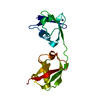
Mass: 190.230 Da / Num. of mol.: 1 / Source method: obtained synthetically / Formula: Os
#3: Water ChemComp-HOH / water


Mass: 18.015 Da / Num. of mol.: 39 / Source method: isolated from a natural source / Formula: H2O

-
Experimental details

-
Experiment

ExperimentMethod: X-RAY DIFFRACTION / Number of used crystals: 1

-
Sample preparation

CrystalDensity Matthews: 2.44 Å3/Da / Density % sol: 47.7 %
Crystal growTemperature: 292 K / Method: vapor diffusion, hanging drop / pH: 8
Details: Tris-HCl buffer, PEG4K, MgCl2, pH 8.0, VAPOR DIFFUSION, HANGING DROP, temperature 292K

-
Data collection

DiffractionMean temperature: 277 K
Diffraction sourceSource: ROTATING ANODE / Type: RIGAKU RU200 / Wavelength: 1.5418 Å
DetectorType: RIGAKU RAXIS IIC / Detector: IMAGE PLATE / Date: May 18, 2001 / Details: mirrors
RadiationProtocol: SINGLE WAVELENGTH / Monochromatic (M) / Laue (L): M / Scattering type: x-ray
Radiation wavelengthWavelength: 1.5418 Å / Relative weight: 1
ReflectionResolution: 2.5→29.55 Å / Num. all: 5624 / Num. obs: 5624 / % possible obs: 93.9 % / Observed criterion σ(F): 1 / Observed criterion σ(I): 2 / Redundancy: 2.5 % / Biso Wilson estimate: 22.3 Å2 / Rsym value: 0.06 / Net I/σ(I): 10.1
Reflection shellResolution: 2.5→2.59 Å / Redundancy: 3.5 % / Mean I/σ(I) obs: 3.7 / Num. unique all: 511 / Rsym value: 0.192 / % possible all: 88.4

-
Processing

Software
NameVersionClassification
CNS1.1refinement
DENZOdata reduction
SCALEPACKdata scaling
CNS1.1phasing
RefinementMethod to determine structure: SIR / Resolution: 2.5→29.55 Å / Rfactor Rfree error: 0.009 / Data cutoff high absF: 142510.37 / Data cutoff low absF: 0 / Isotropic thermal model: RESTRAINED / Cross valid method: THROUGHOUT / σ(F): 0 / Stereochemistry target values: Engh & Huber / Details: BULK SOLVENT MODEL USED
RfactorNum. reflection% reflectionSelection details
Rfree0.223 603 10.7 %RANDOM
Rwork0.206 ---
obs0.206 5624 93.9 %-
all-5624 --
Solvent computationSolvent model: FLAT MODEL / Bsol: 56.9459 Å2 / ksol: 0.37215 e/Å3
Displacement parametersBiso mean: 30.6 Å2
Baniso -1Baniso -2Baniso -3
1-0 Å20 Å20 Å2
2--0 Å20 Å2
3---0 Å2
Refine analyze
FreeObs
Luzzati coordinate error0.34 Å0.29 Å
Luzzati d res low-5 Å
Luzzati sigma a0.36 Å0.32 Å
Refinement stepCycle: LAST / Resolution: 2.5→29.55 Å
ProteinNucleic acidLigandSolventTotal
Num. atoms1173 0 1 39 1213
Refine LS restraints
Refine-IDTypeDev idealDev ideal target
X-RAY DIFFRACTIONc_bond_d0.007
X-RAY DIFFRACTIONc_angle_deg1.3
X-RAY DIFFRACTIONc_dihedral_angle_d24.7
X-RAY DIFFRACTIONc_improper_angle_d0.86
X-RAY DIFFRACTIONc_mcbond_it1.481.5
X-RAY DIFFRACTIONc_mcangle_it2.592
X-RAY DIFFRACTIONc_scbond_it2.122
X-RAY DIFFRACTIONc_scangle_it3.312.5
LS refinement shellResolution: 2.5→2.59 Å / Rfactor Rfree error: 0.045 / Total num. of bins used: 10
RfactorNum. reflection% reflection
Rfree0.336 57 11.2 %
Rwork0.291 454 -
obs-511 88.4 %
Xplor file
Refine-IDSerial noParam fileTopol file
X-RAY DIFFRACTION1PROTEIN_REP.PARAMPROTEIN.TOP
X-RAY DIFFRACTION2WATER.PARAMWATER.TOP
X-RAY DIFFRACTION3ION.PARAMION.TOP

+
About Yorodumi

-
News

-
Feb 9, 2022. New format data for meta-information of EMDB entries

New format data for meta-information of EMDB entries

  • Version 3 of the EMDB header file is now the official format.
  • The previous official version 1.9 will be removed from the archive.

Related info.:EMDB header

External links:wwPDB to switch to version 3 of the EMDB data model

-
Aug 12, 2020. Covid-19 info

Covid-19 info

URL: https://pdbj.org/emnavi/covid19.php

New page: Covid-19 featured information page in EM Navigator.

Related info.:Covid-19 info / Mar 5, 2020. Novel coronavirus structure data

+
Mar 5, 2020. Novel coronavirus structure data

Novel coronavirus structure data

Related info.:Yorodumi Speices / Aug 12, 2020. Covid-19 info

External links:COVID-19 featured content - PDBj / Molecule of the Month (242):Coronavirus Proteases

+
Jan 31, 2019. EMDB accession codes are about to change! (news from PDBe EMDB page)

EMDB accession codes are about to change! (news from PDBe EMDB page)

  • The allocation of 4 digits for EMDB accession codes will soon come to an end. Whilst these codes will remain in use, new EMDB accession codes will include an additional digit and will expand incrementally as the available range of codes is exhausted. The current 4-digit format prefixed with “EMD-” (i.e. EMD-XXXX) will advance to a 5-digit format (i.e. EMD-XXXXX), and so on. It is currently estimated that the 4-digit codes will be depleted around Spring 2019, at which point the 5-digit format will come into force.
  • The EM Navigator/Yorodumi systems omit the EMD- prefix.

Related info.:Q: What is EMD? / ID/Accession-code notation in Yorodumi/EM Navigator

External links:EMDB Accession Codes are Changing Soon! / Contact to PDBj

+
Jul 12, 2017. Major update of PDB

Major update of PDB

  • wwPDB released updated PDB data conforming to the new PDBx/mmCIF dictionary.
  • This is a major update changing the version number from 4 to 5, and with Remediation, in which all the entries are updated.
  • In this update, many items about electron microscopy experimental information are reorganized (e.g. em_software).
  • Now, EM Navigator and Yorodumi are based on the updated data.

External links:wwPDB Remediation / Enriched Model Files Conforming to OneDep Data Standards Now Available in the PDB FTP Archive

-
Yorodumi

Thousand views of thousand structures

  • Yorodumi is a browser for structure data from EMDB, PDB, SASBDB, etc.
  • This page is also the successor to EM Navigator detail page, and also detail information page/front-end page for Omokage search.
  • The word "yorodu" (or yorozu) is an old Japanese word meaning "ten thousand". "mi" (miru) is to see.

Related info.:EMDB / PDB / SASBDB / Comparison of 3 databanks / Yorodumi Search / Aug 31, 2016. New EM Navigator & Yorodumi / Yorodumi Papers / Jmol/JSmol / Function and homology information / Changes in new EM Navigator and Yorodumi

Read more