 Movie
Movie Controller

[English] 日本語
 Yorodumi
- PDB-1z21: Crystal structure of the core domain of Yersinia pestis virulence... -
+ Open data

- Basic information
| Entry | Database: PDB / ID: 1z21 | ||||||
|---|---|---|---|---|---|---|---|
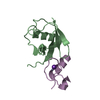
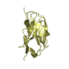
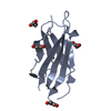
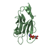
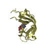
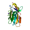
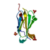
| Title | Crystal structure of the core domain of Yersinia pestis virulence factor YopR | ||||||
 Components Components | Yop proteins translocation protein H | ||||||
 Keywords Keywords | CELL INVASION / Yersinia pestis / plague / type III secretion / YopR / yop | ||||||
| Function / homology |  Function and homology information Function and homology informationtype III protein secretion system complex / protein secretion by the type III secretion system / extracellular region Similarity search - Function | ||||||
| Biological species |   Yersinia pestis (bacteria) Yersinia pestis (bacteria) | ||||||
| Method |  X-RAY DIFFRACTION / SYNCHROTRON / MIR / Resolution: 1.499 Å X-RAY DIFFRACTION / SYNCHROTRON / MIR / Resolution: 1.499 Å | ||||||
 Authors Authors | Schubot, F.D. / Cherry, S. / Tropea, J.E. / Austin, B.P. / Waugh, D.S. | ||||||
 Citation Citation | Journal: Protein Sci. / Year: 2005 Title: Crystal structure of the protease-resistant core domain of Yersinia pestis virulence factor YopR. Authors: Schubot, F.D. / Cherry, S. / Austin, B.P. / Tropea, J.E. / Waugh, D.S. | ||||||
| History |
|
- Structure visualization
| Structure viewer | Molecule: MolmilJmol/JSmol |
|---|
- Downloads & links
-Download
| PDBx/mmCIF format | 1z21.cif.gz | 34.4 KB | Display | PDBx/mmCIF format |
|---|---|---|---|---|
| PDB format | pdb1z21.ent.gz | 23.5 KB | Display | PDB format |
| PDBx/mmJSON format | 1z21.json.gz | Tree view | PDBx/mmJSON format | |
| Others |  Other downloads Other downloads |
-Validation report
| Arichive directory | https://data.pdbj.org/pub/pdb/validation_reports/z2/1z21ftp://data.pdbj.org/pub/pdb/validation_reports/z2/1z21 | HTTPS FTP |
|---|
-Related structure data
| Similar structure data |
|---|


-Links
 PDBj
PDBj- Assembly
| Deposited unit | 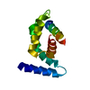
| |||||||||||||||
|---|---|---|---|---|---|---|---|---|---|---|---|---|---|---|---|---|
| 1 |
| |||||||||||||||
| Unit cell |
| |||||||||||||||
| Components on special symmetry positions |
|
-Components
| #1: Protein | Mass: 12606.379 Da / Num. of mol.: 1 / Fragment: YopR(38-149) Source method: isolated from a genetically manipulated source Source: (gene. exp.) Yersinia pestis (bacteria) / Gene: yscH, lcrP / Plasmid: pDEST-MBP / Production host: |
|---|---|
| #2: Water | ChemComp-HOH /  Mass: 18.015 Da / Num. of mol.: 118 / Source method: isolated from a natural source / Formula: H2O Mass: 18.015 Da / Num. of mol.: 118 / Source method: isolated from a natural source / Formula: H2O |
-Experimental details
-Experiment
| Experiment | Method: X-RAY DIFFRACTION / Number of used crystals: 1 |
|---|
- Sample preparation
| Crystal | Density Matthews: 2.1 Å3/Da / Density % sol: 40.3 % |
|---|---|
| Crystal grow | Temperature: 291 K / Method: vapor diffusion, sitting drop / pH: 4.2 Details: 17% PEG-8000, 85 mM Phosphate-citrate, 0.17 M NaCl, and 15% glycerol, pH 4.2, VAPOR DIFFUSION, SITTING DROP, temperature 291K |
-Data collection
| Diffraction | Mean temperature: 100 K |
|---|---|
| Diffraction source | Source: SYNCHROTRON / Site: NSLS  / Beamline: X9B / Wavelength: 1 Å / Beamline: X9B / Wavelength: 1 Å |
| Detector | Type: BRANDEIS / Detector: CCD / Date: Sep 15, 2003 |
| Radiation | Protocol: SINGLE WAVELENGTH / Monochromatic (M) / Laue (L): M / Scattering type: x-ray |
| Radiation wavelength | Wavelength: 1 Å / Relative weight: 1 |
| Reflection | Resolution: 1.5→22.82 Å / Num. all: 17699 / Num. obs: 17807 / % possible obs: 99.3 % / Observed criterion σ(F): 2 / Observed criterion σ(I): 0 / Redundancy: 4.5 % / Rmerge(I) obs: 0.036 / Rsym value: 0.036 / Net I/σ(I): 46.1 |
| Reflection shell | Resolution: 1.5→1.54 Å / % possible all: 97.5 |
- Processing
| Software |
| ||||||||||||||||||||||||||||||||||||||||||||||||||||||||||||||||||||||||||||||||||||||||||||||||||||
|---|---|---|---|---|---|---|---|---|---|---|---|---|---|---|---|---|---|---|---|---|---|---|---|---|---|---|---|---|---|---|---|---|---|---|---|---|---|---|---|---|---|---|---|---|---|---|---|---|---|---|---|---|---|---|---|---|---|---|---|---|---|---|---|---|---|---|---|---|---|---|---|---|---|---|---|---|---|---|---|---|---|---|---|---|---|---|---|---|---|---|---|---|---|---|---|---|---|---|---|---|---|
| Refinement | Method to determine structure: MIR / Resolution: 1.499→22.81 Å / Cor.coef. Fo:Fc: 0.953 / Cor.coef. Fo:Fc free: 0.936 / SU B: 2.544 / SU ML: 0.082 / Cross valid method: THROUGHOUT / σ(F): 2 / ESU R: 0.112 / ESU R Free: 0.11 / Stereochemistry target values: MAXIMUM LIKELIHOOD / Details: HYDROGENS HAVE BEEN ADDED IN THE RIDING POSITIONS
| ||||||||||||||||||||||||||||||||||||||||||||||||||||||||||||||||||||||||||||||||||||||||||||||||||||
| Solvent computation | Ion probe radii: 0.8 Å / Shrinkage radii: 0.8 Å / VDW probe radii: 1.4 Å / Solvent model: BABINET MODEL WITH MASK | ||||||||||||||||||||||||||||||||||||||||||||||||||||||||||||||||||||||||||||||||||||||||||||||||||||
| Displacement parameters | Biso mean: 21.596 Å2 | ||||||||||||||||||||||||||||||||||||||||||||||||||||||||||||||||||||||||||||||||||||||||||||||||||||
| Refinement step | Cycle: LAST / Resolution: 1.499→22.81 Å
| ||||||||||||||||||||||||||||||||||||||||||||||||||||||||||||||||||||||||||||||||||||||||||||||||||||
| Refine LS restraints |
| ||||||||||||||||||||||||||||||||||||||||||||||||||||||||||||||||||||||||||||||||||||||||||||||||||||
| LS refinement shell | Resolution: 1.5→1.539 Å / Total num. of bins used: 20 /
|
