- PDB-1yyp: Crystal structure of cytomegalovirus UL44 bound to C-terminal pep... -
+
Open data
ID or keywords:
Loading...
-
Basic information
Entry
Database: PDB / ID: 1yyp
Title
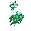
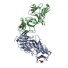
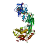
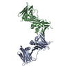
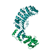
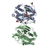
Crystal structure of cytomegalovirus UL44 bound to C-terminal peptide from CMV UL54
Components
DNA polymerase
DNA polymerase processivity factor
Keywords
REPLICATION/TRANSFERASE / PROCESSIVITY FOLD (SAME FOLD AS HSV UL42 / PCNA / AND HOMOTRIMERIC SLIDING CLAMPS) / REPLICATION-TRANSFERASE COMPLEX
Function / homology
Function and homology information
bidirectional double-stranded viral DNA replication / DNA polymerase processivity factor activity / DNA-templated DNA replication / virion component / DNA-directed DNA polymerase / symbiont-mediated suppression of host cytoplasmic pattern recognition receptor signaling pathway via inhibition of IRF3 activity / symbiont-mediated suppression of host NF-kappaB cascade / DNA-directed DNA polymerase activity / DNA replication / nucleotide binding ...bidirectional double-stranded viral DNA replication / DNA polymerase processivity factor activity / DNA-templated DNA replication / virion component / DNA-directed DNA polymerase / symbiont-mediated suppression of host cytoplasmic pattern recognition receptor signaling pathway via inhibition of IRF3 activity / symbiont-mediated suppression of host NF-kappaB cascade / DNA-directed DNA polymerase activity / DNA replication / nucleotide binding / host cell nucleus / DNA binding Similarity search - Function
Herpesvirus polymerase accessory protein / Herpesvirus polymerase accessory protein / Box / Proliferating Cell Nuclear Antigen / Proliferating Cell Nuclear Antigen - #10 / : / DNA polymerase family B, thumb domain / DNA-directed DNA polymerase, family B, multifunctional domain / DNA-directed DNA polymerase, family B, conserved site / DNA polymerase family B signature. ...Herpesvirus polymerase accessory protein / Herpesvirus polymerase accessory protein / Box / Proliferating Cell Nuclear Antigen / Proliferating Cell Nuclear Antigen - #10 / : / DNA polymerase family B, thumb domain / DNA-directed DNA polymerase, family B, multifunctional domain / DNA-directed DNA polymerase, family B, conserved site / DNA polymerase family B signature. / DNA polymerase family B / DNA polymerase family B, exonuclease domain / DNA-directed DNA polymerase, family B, exonuclease domain / DNA polymerase, palm domain superfamily / DNA polymerase type-B family / DNA-directed DNA polymerase, family B / : / Ribonuclease H superfamily / Ribonuclease H-like superfamily / DNA/RNA polymerase superfamily / Alpha Beta Similarity search - Domain/homology
#2: Journal: J.Virol. / Year: 2004 Title: Specific residues in the connector loop of the human cytomegalovirus DNA polymerase accessory protein UL44 are crucial for interaction with the UL54 catalytic subunit
#3: Journal: J.Virol. / Year: 2004 Title: Residues of human cytomegalovirus DNA polymerase catalytic subunit UL54 that are necessary and sufficient for interaction with the accessory protein UL44
#4: Journal: J.Virol. / Year: 1992 Title: Physical and functional interaction of human cytomegalovirus DNA polymerase and its accessory protein (ICP36) expressed in insect cells
History
Deposition
Feb 25, 2005
Deposition site: RCSB / Processing site: RCSB
Revision 1.0
Dec 27, 2005
Provider: repository / Type: Initial release
Revision 1.1
Apr 30, 2008
Group: Version format compliance
Revision 1.2
Jul 13, 2011
Group: Advisory / Refinement description / Version format compliance
SEQUENCE AUTHOR STATES THAT THE SER 205 IS INDEED CORRECT AND IT MAY REPRESENT A DIFFERENT ISOLATE ...SEQUENCE AUTHOR STATES THAT THE SER 205 IS INDEED CORRECT AND IT MAY REPRESENT A DIFFERENT ISOLATE CYTOMEGALOVIRUS, REFERRING TO ERTL PF, POWELL KL. "PHYSICAL AND FUNCTIONAL INTERACTION OF HUMAN CYTOMEGALOVIRUS DNA POLYMERASE AND ITS ACCESSORY PROTEIN (ICP36) EXPRESSED IN INSECT CELLS." J VIROL. 1992 JUL;66(7):4126-33.
Mass: 18.015 Da / Num. of mol.: 48 / Source method: isolated from a natural source / Formula: H2O
-
Experimental details
-
Experiment
Experiment
Method: X-RAY DIFFRACTION / Number of used crystals: 1
-
Sample preparation
Crystal
Density Matthews: 2.8 Å3/Da / Density % sol: 55 %
Crystal grow
Temperature: 295 K / Method: vapor diffusion, hanging drop / pH: 4.2 Details: 2 M ammonium sulfate, 100 mM phosphate-citrate, and 10 mM DTT , pH 4.2, VAPOR DIFFUSION, HANGING DROP, temperature 295K
In the structure databanks used in Yorodumi, some data are registered as the other names, "COVID-19 virus" and "2019-nCoV". Here are the details of the virus and the list of structure data.
Jan 31, 2019. EMDB accession codes are about to change! (news from PDBe EMDB page)
EMDB accession codes are about to change! (news from PDBe EMDB page)
The allocation of 4 digits for EMDB accession codes will soon come to an end. Whilst these codes will remain in use, new EMDB accession codes will include an additional digit and will expand incrementally as the available range of codes is exhausted. The current 4-digit format prefixed with “EMD-” (i.e. EMD-XXXX) will advance to a 5-digit format (i.e. EMD-XXXXX), and so on. It is currently estimated that the 4-digit codes will be depleted around Spring 2019, at which point the 5-digit format will come into force.
The EM Navigator/Yorodumi systems omit the EMD- prefix.
Related info.:Q: What is EMD? / ID/Accession-code notation in Yorodumi/EM Navigator
Yorodumi is a browser for structure data from EMDB, PDB, SASBDB, etc.
This page is also the successor to EM Navigator detail page, and also detail information page/front-end page for Omokage search.
The word "yorodu" (or yorozu) is an old Japanese word meaning "ten thousand". "mi" (miru) is to see.
Related info.:EMDB / PDB / SASBDB / Comparison of 3 databanks / Yorodumi Search / Aug 31, 2016. New EM Navigator & Yorodumi / Yorodumi Papers / Jmol/JSmol / Function and homology information / Changes in new EM Navigator and Yorodumi
 Movie
Movie Controller

 Yorodumi
 Open data
 Basic information
 Components
 Keywords
 Function and homology information
 Human herpesvirus 5
 X-RAY DIFFRACTION /
  Authors
 Citation
 Structure visualization
 Downloads & links
 Other downloads


 PDBj
PDBj


 Assembly
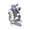


 Mass: 96.063 Da / Num. of mol.: 4 / Source method: obtained synthetically / Formula: SO4
 Mass: 62.068 Da / Num. of mol.: 3 / Source method: obtained synthetically / Formula: C2H6O2
 Mass: 18.015 Da / Num. of mol.: 48 / Source method: isolated from a natural source / Formula: H2O
 Sample preparation
 / Beamline: 19-ID / Wavelength: 0.9793, 0.9794, 1.008, 0.9537
 Processing