 Movie
Movie Controller

[English] 日本語
 Yorodumi
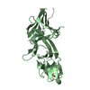
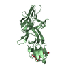
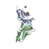
- PDB-3m7j: Crystal structure of the bacteriocin LLPA from pseudomonas sp. in... -
+ Open data

- Basic information
| Entry | Database: PDB / ID: 3m7j | ||||||
|---|---|---|---|---|---|---|---|
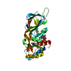
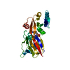
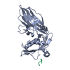
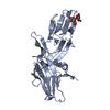
| Title | Crystal structure of the bacteriocin LLPA from pseudomonas sp. in complex with Met-mannose | ||||||
 Components Components | Putidacin L1 | ||||||
 Keywords Keywords | ANTIMICROBIAL PROTEIN / monocot mannose-binding lectin / bacteriocin / LLPA / Pseudomonas / bacterial toxin / SIRAS / protein-sugar complex / mannose | ||||||
| Function / homology |  Function and homology information Function and homology informationAgglutinin, subunit A / Bulb-type lectin domain / Bulb-type lectin domain / Bulb-type lectin domain superfamily / Bulb-type lectin domain profile. / Bulb-type mannose-specific lectin / Orthogonal Prism / Mainly Beta Similarity search - Domain/homology | ||||||
| Biological species |  Pseudomonas sp. (bacteria) Pseudomonas sp. (bacteria) | ||||||
| Method |  X-RAY DIFFRACTION / SYNCHROTRON / MOLECULAR REPLACEMENT / Resolution: 2.26 Å X-RAY DIFFRACTION / SYNCHROTRON / MOLECULAR REPLACEMENT / Resolution: 2.26 Å | ||||||
 Authors Authors | Loris, R. / Garcia-Pino, A. | ||||||
 Citation Citation | Journal: Plos Pathog. / Year: 2013 Title: Structural Determinants for Activity and Specificity of the Bacterial Toxin LlpA Authors: Ghequire, M.G. / Garcia-Pino, A. / Lebbe, E.K. / Spaepen, S. / Loris, R. / De Mot, R. | ||||||
| History |
|
- Structure visualization
| Structure viewer | Molecule: MolmilJmol/JSmol |
|---|
- Downloads & links
-Download
| PDBx/mmCIF format | 3m7j.cif.gz | 208.4 KB | Display | PDBx/mmCIF format |
|---|---|---|---|---|
| PDB format | pdb3m7j.ent.gz | 167.4 KB | Display | PDB format |
| PDBx/mmJSON format | 3m7j.json.gz | Tree view | PDBx/mmJSON format | |
| Others |  Other downloads Other downloads |
-Validation report
| Arichive directory | https://data.pdbj.org/pub/pdb/validation_reports/m7/3m7jftp://data.pdbj.org/pub/pdb/validation_reports/m7/3m7j | HTTPS FTP |
|---|
-Related structure data
| Related structure data | 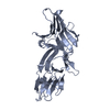 3m7hSC  4gc1C 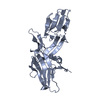 4gc2C S: Starting model for refinement C: citing same article ( |
|---|---|
| Similar structure data |


-Links
 PDBj
PDBj
- Assembly
| Deposited unit | 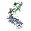
| ||||||||
|---|---|---|---|---|---|---|---|---|---|
| 1 | 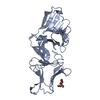
| ||||||||
| 2 | 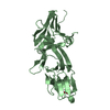
| ||||||||
| Unit cell |
|
-Components
| #1: Protein | Mass: 29993.719 Da / Num. of mol.: 2 Source method: isolated from a genetically manipulated source Source: (gene. exp.) Pseudomonas sp. (bacteria) / Strain: BW11M1 / Gene: llpa / Production host: #2: Sugar | 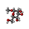  Type: D-saccharide / Mass: 194.182 Da / Num. of mol.: 2 Type: D-saccharide / Mass: 194.182 Da / Num. of mol.: 2Source method: isolated from a genetically manipulated source Formula: C7H14O6 #3: Water | ChemComp-HOH / |  Mass: 18.015 Da / Num. of mol.: 113 / Source method: isolated from a natural source / Formula: H2O Mass: 18.015 Da / Num. of mol.: 113 / Source method: isolated from a natural source / Formula: H2O |
|---|
-Experimental details
-Experiment
| Experiment | Method: X-RAY DIFFRACTION / Number of used crystals: 1 |
|---|
- Sample preparation
| Crystal | Density Matthews: 3.23 Å3/Da / Density % sol: 61.87 % |
|---|---|
| Crystal grow | Temperature: 293 K / Method: vapor diffusion, hanging drop / pH: 6.5 Details: 0.1M imidazole, 1.3M sodium acetate, pH 6.5, VAPOR DIFFUSION, HANGING DROP, temperature 293K |
-Data collection
| Diffraction | Mean temperature: 293 K |
|---|---|
| Diffraction source | Source: SYNCHROTRON / Site: EMBL/DESY, HAMBURG  / Beamline: X13 / Wavelength: 0.8073 Å / Beamline: X13 / Wavelength: 0.8073 Å |
| Detector | Type: MAR CCD 165 mm / Detector: CCD / Date: May 10, 2008 / Details: mirrors |
| Radiation | Protocol: SINGLE WAVELENGTH / Monochromatic (M) / Laue (L): M / Scattering type: x-ray |
| Radiation wavelength | Wavelength: 0.8073 Å / Relative weight: 1 |
| Reflection | Resolution: 2.26→19.645 Å / Num. all: 33688 / Num. obs: 33688 / % possible obs: 100 % / Observed criterion σ(F): 0 / Observed criterion σ(I): 0 / Biso Wilson estimate: 34.33 Å2 / Rmerge(I) obs: 0.124 / Rsym value: 0.124 / Net I/σ(I): 7.1 |
| Reflection shell | Resolution: 2.26→2.34 Å / Rmerge(I) obs: 0.542 / Mean I/σ(I) obs: 2.6 / Num. unique all: 3360 / Rsym value: 0.542 / % possible all: 100 |
- Processing
| Software |
| |||||||||||||||||||||||||||||||||||||||||||||||||||||||||||||||||||||||||||||||||||||||||||||||||||||||||||||||||||||||||||||||||||||
|---|---|---|---|---|---|---|---|---|---|---|---|---|---|---|---|---|---|---|---|---|---|---|---|---|---|---|---|---|---|---|---|---|---|---|---|---|---|---|---|---|---|---|---|---|---|---|---|---|---|---|---|---|---|---|---|---|---|---|---|---|---|---|---|---|---|---|---|---|---|---|---|---|---|---|---|---|---|---|---|---|---|---|---|---|---|---|---|---|---|---|---|---|---|---|---|---|---|---|---|---|---|---|---|---|---|---|---|---|---|---|---|---|---|---|---|---|---|---|---|---|---|---|---|---|---|---|---|---|---|---|---|---|---|---|
| Refinement | Method to determine structure: MOLECULAR REPLACEMENT Starting model: PDB ENTRY 3M7H Resolution: 2.26→19.644 Å / Occupancy max: 1 / Occupancy min: 1 / FOM work R set: 0.847 / SU ML: 1.72 / Cross valid method: THROUGHOUT / σ(F): 0.05 / σ(I): 0 / Phase error: 21.83 / Stereochemistry target values: ML
| |||||||||||||||||||||||||||||||||||||||||||||||||||||||||||||||||||||||||||||||||||||||||||||||||||||||||||||||||||||||||||||||||||||
| Solvent computation | Shrinkage radii: 0.9 Å / VDW probe radii: 1.11 Å / Solvent model: FLAT BULK SOLVENT MODEL / Bsol: 50.697 Å2 / ksol: 0.335 e/Å3 | |||||||||||||||||||||||||||||||||||||||||||||||||||||||||||||||||||||||||||||||||||||||||||||||||||||||||||||||||||||||||||||||||||||
| Displacement parameters | Biso max: 104.43 Å2 / Biso mean: 44.515 Å2 / Biso min: 17.55 Å2
| |||||||||||||||||||||||||||||||||||||||||||||||||||||||||||||||||||||||||||||||||||||||||||||||||||||||||||||||||||||||||||||||||||||
| Refinement step | Cycle: LAST / Resolution: 2.26→19.644 Å
| |||||||||||||||||||||||||||||||||||||||||||||||||||||||||||||||||||||||||||||||||||||||||||||||||||||||||||||||||||||||||||||||||||||
| Refine LS restraints |
| |||||||||||||||||||||||||||||||||||||||||||||||||||||||||||||||||||||||||||||||||||||||||||||||||||||||||||||||||||||||||||||||||||||
| LS refinement shell |
| |||||||||||||||||||||||||||||||||||||||||||||||||||||||||||||||||||||||||||||||||||||||||||||||||||||||||||||||||||||||||||||||||||||
| Refinement TLS params. | Method: refined / Refine-ID: X-RAY DIFFRACTION
| |||||||||||||||||||||||||||||||||||||||||||||||||||||||||||||||||||||||||||||||||||||||||||||||||||||||||||||||||||||||||||||||||||||
| Refinement TLS group |
|
