 Movie
Movie Controller

[English] 日本語
 Yorodumi
- PDB-1yxy: Crystal Structure of putative N-acetylmannosamine-6-P epimerase f... -
+ Open data

- Basic information
| Entry | Database: PDB / ID: 1yxy | ||||||
|---|---|---|---|---|---|---|---|
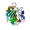
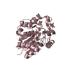
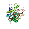
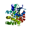
| Title | Crystal Structure of putative N-acetylmannosamine-6-P epimerase from Streptococcus pyogenes (APC29713) Structural genomics, MCSG | ||||||
 Components Components | Putative N-acetylmannosamine-6-phosphate 2-epimerase | ||||||
 Keywords Keywords | ISOMERASE / Structural genomics / epimerase / Streptococcus pyogenes / PSI / Protein Structure Initiative / Midwest Center for Structural Genomics / MCSG | ||||||
| Function / homology |  Function and homology information Function and homology informationN-acylglucosamine-6-phosphate 2-epimerase / N-acetylmannosamine catabolic process / N-acylglucosamine-6-phosphate 2-epimerase activity / N-acetylneuraminate catabolic process / carbohydrate metabolic process / cytosol Similarity search - Function | ||||||
| Biological species |  Streptococcus pyogenes (bacteria) Streptococcus pyogenes (bacteria) | ||||||
| Method |  X-RAY DIFFRACTION / SYNCHROTRON / SAD / Resolution: 1.6 Å X-RAY DIFFRACTION / SYNCHROTRON / SAD / Resolution: 1.6 Å | ||||||
 Authors Authors | Rotella, F.J. / Zhang, R.G. / Lezondra, L.E.O. / Collart, F.R. / Joachimiak, A. / Midwest Center for Structural Genomics (MCSG) | ||||||
 Citation Citation | Journal: To be Published Title: The 1.6 A crystal structure of putative N-acetylmannosamine-6-P epimerase from Streptococcus pyogenes Authors: Rotella, F.J. / Zhang, R.G. / Lezondra, L.E.O. / Collart, F.R. / Joachimiak, A. | ||||||
| History |
|
- Structure visualization
| Structure viewer | Molecule: MolmilJmol/JSmol |
|---|
- Downloads & links
-Download
| PDBx/mmCIF format | 1yxy.cif.gz | 111 KB | Display | PDBx/mmCIF format |
|---|---|---|---|---|
| PDB format | pdb1yxy.ent.gz | 85.9 KB | Display | PDB format |
| PDBx/mmJSON format | 1yxy.json.gz | Tree view | PDBx/mmJSON format | |
| Others |  Other downloads Other downloads |
-Validation report
| Arichive directory | https://data.pdbj.org/pub/pdb/validation_reports/yx/1yxyftp://data.pdbj.org/pub/pdb/validation_reports/yx/1yxy | HTTPS FTP |
|---|
-Related structure data
| Similar structure data | |
|---|---|
| Other databases |


-Links
 PDBj
PDBj
- Assembly
| Deposited unit | 
| ||||||||
|---|---|---|---|---|---|---|---|---|---|
| 1 | 
| ||||||||
| 2 | 
| ||||||||
| 3 | 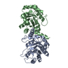
| ||||||||
| 4 |
| ||||||||
| Unit cell |
| ||||||||
| Details | the biological assembly is a dimer |
-Components
| #1: Protein | Mass: 25094.020 Da / Num. of mol.: 2 Source method: isolated from a genetically manipulated source Source: (gene. exp.) Streptococcus pyogenes (bacteria) / Gene: nanE / Plasmid: PDM68 / Species (production host): Escherichia coli / Production host: References: UniProt: P65522, N-acylglucosamine-6-phosphate 2-epimerase #2: Water | ChemComp-HOH / |  Mass: 18.015 Da / Num. of mol.: 679 / Source method: isolated from a natural source / Formula: H2O Mass: 18.015 Da / Num. of mol.: 679 / Source method: isolated from a natural source / Formula: H2O |
|---|
-Experimental details
-Experiment
| Experiment | Method: X-RAY DIFFRACTION / Number of used crystals: 1 |
|---|
- Sample preparation
| Crystal | Density Matthews: 2.69 Å3/Da / Density % sol: 53.86 % |
|---|---|
| Crystal grow | Temperature: 273 K / Method: vapor diffusion, sitting drop / pH: 6.5 Details: ammonium sulfate, BIS-TRIS buffer, pH 6.5, VAPOR DIFFUSION, SITTING DROP, temperature 273K |
-Data collection
| Diffraction | Mean temperature: 100 K |
|---|---|
| Diffraction source | Source: SYNCHROTRON / Site: APS  / Beamline: 19-BM / Wavelength: 0.979484 Å / Beamline: 19-BM / Wavelength: 0.979484 Å |
| Detector | Type: SBC-3 / Detector: CCD / Date: Nov 7, 2004 Details: horizontal-focussing Si-111 double-crystal monochromator and vertical-focusing mirror |
| Radiation | Monochromator: sagitally focused Si-111 / Protocol: SINGLE WAVELENGTH / Monochromatic (M) / Laue (L): M / Scattering type: x-ray |
| Radiation wavelength | Wavelength: 0.979484 Å / Relative weight: 1 |
| Reflection | Resolution: 1.6→50 Å / Num. all: 69461 / Num. obs: 68381 / % possible obs: 98.4 % / Observed criterion σ(F): 1 / Observed criterion σ(I): 1 / Redundancy: 13 % / Biso Wilson estimate: 30.5 Å2 / Rmerge(I) obs: 0.123 / Net I/σ(I): 26 |
| Reflection shell | Resolution: 1.6→1.66 Å / Redundancy: 10.1 % / Rmerge(I) obs: 0.809 / Mean I/σ(I) obs: 2.4 / Num. unique all: 6793 / % possible all: 98.2 |
- Processing
| Software |
| ||||||||||||||||||||||||||||||||||||||||||||||||||||||||||||||||||||||||||||||||||||||||||
|---|---|---|---|---|---|---|---|---|---|---|---|---|---|---|---|---|---|---|---|---|---|---|---|---|---|---|---|---|---|---|---|---|---|---|---|---|---|---|---|---|---|---|---|---|---|---|---|---|---|---|---|---|---|---|---|---|---|---|---|---|---|---|---|---|---|---|---|---|---|---|---|---|---|---|---|---|---|---|---|---|---|---|---|---|---|---|---|---|---|---|---|
| Refinement | Method to determine structure: SAD / Resolution: 1.6→50 Å / Cor.coef. Fo:Fc: 0.959 / Cor.coef. Fo:Fc free: 0.954 / SU B: 1.531 / SU ML: 0.055 / Isotropic thermal model: RESTRAINED / Cross valid method: THROUGHOUT / σ(F): 0 / ESU R: 0.091 / ESU R Free: 0.084 Stereochemistry target values: MAXIMUM LIKELIHOOD WITH PHASES Details: HYDROGENS HAVE BEEN ADDED IN THE RIDING POSITIONS
| ||||||||||||||||||||||||||||||||||||||||||||||||||||||||||||||||||||||||||||||||||||||||||
| Solvent computation | Ion probe radii: 0.8 Å / Shrinkage radii: 0.8 Å / VDW probe radii: 1.2 Å / Solvent model: MASK | ||||||||||||||||||||||||||||||||||||||||||||||||||||||||||||||||||||||||||||||||||||||||||
| Displacement parameters | Biso mean: 22.286 Å2
| ||||||||||||||||||||||||||||||||||||||||||||||||||||||||||||||||||||||||||||||||||||||||||
| Refine analyze | Luzzati coordinate error obs: 0.182 Å | ||||||||||||||||||||||||||||||||||||||||||||||||||||||||||||||||||||||||||||||||||||||||||
| Refinement step | Cycle: LAST / Resolution: 1.6→50 Å
| ||||||||||||||||||||||||||||||||||||||||||||||||||||||||||||||||||||||||||||||||||||||||||
| Refine LS restraints |
| ||||||||||||||||||||||||||||||||||||||||||||||||||||||||||||||||||||||||||||||||||||||||||
| LS refinement shell | Resolution: 1.6→1.639 Å / Total num. of bins used: 20
|
