 Movie
Movie Controller

[English] 日本語
 Yorodumi
- PDB-1yo6: Crystal Structure of the putative Carbonyl Reductase Sniffer of C... -
+ Open data

- Basic information
| Entry | Database: PDB / ID: 1yo6 | ||||||
|---|---|---|---|---|---|---|---|
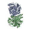
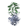
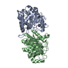
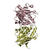
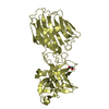
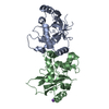
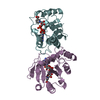
| Title | Crystal Structure of the putative Carbonyl Reductase Sniffer of Caenorhabditis elegans | ||||||
 Components Components | Putative Carbonyl Reductase Sniffer | ||||||
 Keywords Keywords | OXIDOREDUCTASE / Tyrosine-dependent oxidoreductase (SDR family) / Structural Genomics / PSI / Protein Structure Initiative / Southeast Collaboratory for Structural Genomics / SECSG | ||||||
| Function / homology |  Function and homology information Function and homology information | ||||||
| Biological species |  | ||||||
| Method |  X-RAY DIFFRACTION / SYNCHROTRON / MOLECULAR REPLACEMENT / Resolution: 2.6 Å X-RAY DIFFRACTION / SYNCHROTRON / MOLECULAR REPLACEMENT / Resolution: 2.6 Å | ||||||
 Authors Authors | Southeast Collaboratory for Structural Genomics (SECSG) | ||||||
 Citation Citation | Journal: To be Published Title: Crystal Structure of the putative Carbonyl Reductase Sniffer of Caenorhabditis elegans Authors: Schormann, N. / Lu, S. / Symersky, J. / Karpova, E. / Zhang, Y. / Luo, D. / Zhou, Q. / Huang, W. / Luan, C.-H. / Gray, R. / Arabshahi, A. / McKinstry, A. / Qiu, S. / Cao, Z. / An, J. / Li, S. ...Authors: Schormann, N. / Lu, S. / Symersky, J. / Karpova, E. / Zhang, Y. / Luo, D. / Zhou, Q. / Huang, W. / Luan, C.-H. / Gray, R. / Arabshahi, A. / McKinstry, A. / Qiu, S. / Cao, Z. / An, J. / Li, S. / Lin, G. / Tsao, J. / Johnson, D. / Luo, M. / Shang, Q. / Chen, Y. / Stinnett, M. / Bunzel, R. / Carson, M. / Bray, T. / DeLucas, L. | ||||||
| History |
|
- Structure visualization
| Structure viewer | Molecule: MolmilJmol/JSmol |
|---|
- Downloads & links
-Download
| PDBx/mmCIF format | 1yo6.cif.gz | 270.2 KB | Display | PDBx/mmCIF format |
|---|---|---|---|---|
| PDB format | pdb1yo6.ent.gz | 220.1 KB | Display | PDB format |
| PDBx/mmJSON format | 1yo6.json.gz | Tree view | PDBx/mmJSON format | |
| Others |  Other downloads Other downloads |
-Validation report
| Arichive directory | https://data.pdbj.org/pub/pdb/validation_reports/yo/1yo6ftp://data.pdbj.org/pub/pdb/validation_reports/yo/1yo6 | HTTPS FTP |
|---|
-Related structure data
| Related structure data | 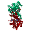 1snyS S: Starting model for refinement |
|---|---|
| Similar structure data | |
| Other databases |


-Links
 PDBj
PDBj

- Assembly
| Deposited unit | 
| ||||||||||
|---|---|---|---|---|---|---|---|---|---|---|---|
| 1 | 
| ||||||||||
| 2 | 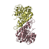
| ||||||||||
| 3 | 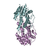
| ||||||||||
| Unit cell |
| ||||||||||
| Details | The biological assembly is a dimer (chains A,B; chains C,D; chains E,F). The asymmetric unit contains 3 independent homodimers. |
-Components
| #1: Protein | Mass: 26730.551 Da / Num. of mol.: 6 Source method: isolated from a genetically manipulated source Details: Apo-Enzyme / Source: (gene. exp.)  #2: Water | ChemComp-HOH / |  Mass: 18.015 Da / Num. of mol.: 236 / Source method: isolated from a natural source / Formula: H2O Mass: 18.015 Da / Num. of mol.: 236 / Source method: isolated from a natural source / Formula: H2O |
|---|
-Experimental details
-Experiment
| Experiment | Method: X-RAY DIFFRACTION / Number of used crystals: 1 |
|---|
- Sample preparation
| Crystal | Density Matthews: 2.4 Å3/Da / Density % sol: 48.8 % |
|---|---|
| Crystal grow | Temperature: 295 K / Method: vapor diffusion, hanging drop / pH: 7.4 Details: 20% PEG2000 MME, 0.1M Tris, 0.1% beta-OG, pH 7.4, VAPOR DIFFUSION, HANGING DROP, temperature 295K |
-Data collection
| Diffraction | Mean temperature: 100 K |
|---|---|
| Diffraction source | Source: SYNCHROTRON / Site: APS  / Beamline: 22-ID / Wavelength: 0.9793 Å / Beamline: 22-ID / Wavelength: 0.9793 Å |
| Detector | Type: MARMOSAIC 225 mm CCD / Detector: CCD / Date: Dec 8, 2004 |
| Radiation | Monochromator: Graphite / Protocol: SINGLE WAVELENGTH / Monochromatic (M) / Laue (L): M / Scattering type: x-ray |
| Radiation wavelength | Wavelength: 0.9793 Å / Relative weight: 1 |
| Reflection | Resolution: 2.6→50 Å / Num. all: 46212 / Num. obs: 46212 / % possible obs: 94.3 % / Observed criterion σ(I): 1 / Redundancy: 4.6 % / Biso Wilson estimate: 45.5 Å2 / Rmerge(I) obs: 0.06 / Rsym value: 0.06 / Net I/σ(I): 19.5 |
| Reflection shell | Resolution: 2.6→2.69 Å / Redundancy: 4.5 % / Rmerge(I) obs: 0.205 / Mean I/σ(I) obs: 4.2 / Num. unique all: 4124 / Rsym value: 0.205 / % possible all: 86.2 |
- Processing
| Software |
| ||||||||||||||||||||||||||||||||||||
|---|---|---|---|---|---|---|---|---|---|---|---|---|---|---|---|---|---|---|---|---|---|---|---|---|---|---|---|---|---|---|---|---|---|---|---|---|---|
| Refinement | Method to determine structure: MOLECULAR REPLACEMENT Starting model: The starting model was created by the SWISS-MODEL server based on PDB entry 1SNY using the C. elegans target sequence (C55A6.5) Resolution: 2.6→47.12 Å / Rfactor Rfree error: 0.006 / Data cutoff high absF: 6442715.26 / Data cutoff low absF: 0 / Isotropic thermal model: RESTRAINED / Cross valid method: THROUGHOUT / σ(F): 0 / Stereochemistry target values: Engh & Huber Details: The following residues are not visible in the electron density: 205-217 chain A; 204-218 chain B; 208-217 chain C; 205-218 chain D; 1,204-217 chain E; 1,48-49,81-82,136-137,206-218 chain F
| ||||||||||||||||||||||||||||||||||||
| Solvent computation | Solvent model: FLAT MODEL / Bsol: 43.5276 Å2 / ksol: 0.336883 e/Å3 | ||||||||||||||||||||||||||||||||||||
| Displacement parameters | Biso mean: 48.1 Å2
| ||||||||||||||||||||||||||||||||||||
| Refine analyze |
| ||||||||||||||||||||||||||||||||||||
| Refinement step | Cycle: LAST / Resolution: 2.6→47.12 Å
| ||||||||||||||||||||||||||||||||||||
| Refine LS restraints |
| ||||||||||||||||||||||||||||||||||||
| LS refinement shell | Resolution: 2.6→2.76 Å / Rfactor Rfree error: 0.021 / Total num. of bins used: 6
| ||||||||||||||||||||||||||||||||||||
| Xplor file |
|
