 Movie
Movie Controller

[English] 日本語
 Yorodumi
- PDB-1yny: Molecular Structure of D-Hydantoinase from a Bacillus sp. AR9: Ev... -
+ Open data

- Basic information
| Entry | Database: PDB / ID: 1yny | ||||||
|---|---|---|---|---|---|---|---|
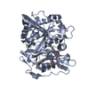
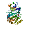
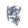
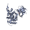
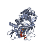
| Title | Molecular Structure of D-Hydantoinase from a Bacillus sp. AR9: Evidence for mercury inhibition | ||||||
 Components Components | D-Hydantoinase | ||||||
 Keywords Keywords | HYDROLASE / TIM-barrel / hydantoinase / binuclear metal-binding | ||||||
| Function / homology |  Function and homology information Function and homology informationdihydropyrimidinase / dihydropyrimidinase activity / metal ion binding / cytosol Similarity search - Function | ||||||
| Biological species |  | ||||||
| Method |  X-RAY DIFFRACTION / MOLECULAR REPLACEMENT / Resolution: 2.3 Å X-RAY DIFFRACTION / MOLECULAR REPLACEMENT / Resolution: 2.3 Å | ||||||
 Authors Authors | Radha Kishan, K.V. / Vohra, R.M. / Ganeshan, K. / Agrawal, V. / Sharma, V.M. / Sharma, R. | ||||||
 Citation Citation | Journal: J.Mol.Biol. / Year: 2005 Title: Molecular structure of D-hydantoinase from Bacillus sp. AR9: evidence for mercury inhibition. Authors: Radha Kishan, K.V. / Vohra, R.M. / Ganesan, K. / Agrawal, V. / Sharma, V.M. / Sharma, R. #1: Journal: Acta Crystallogr.,Sect.D / Year: 2002Title: Crystallization and preliminary X-ray diffraction analysis of a thermostable D-hydantoinase from mesophilic Bacillus sp.AR9 Authors: Agrawal, V. / Sharma, R. / Ganeshan, K. / Radha Kishan, K.V. | ||||||
| History |
|
- Structure visualization
| Structure viewer | Molecule: MolmilJmol/JSmol |
|---|
- Downloads & links
-Download
| PDBx/mmCIF format | 1yny.cif.gz | 189.4 KB | Display | PDBx/mmCIF format |
|---|---|---|---|---|
| PDB format | pdb1yny.ent.gz | 150 KB | Display | PDB format |
| PDBx/mmJSON format | 1yny.json.gz | Tree view | PDBx/mmJSON format | |
| Others |  Other downloads Other downloads |
-Validation report
| Arichive directory | https://data.pdbj.org/pub/pdb/validation_reports/yn/1ynyftp://data.pdbj.org/pub/pdb/validation_reports/yn/1yny | HTTPS FTP |
|---|
-Related structure data
| Related structure data |  1gkpS S: Starting model for refinement |
|---|---|
| Similar structure data |


-Links
 PDBj
PDBj

- Assembly
| Deposited unit | 
| ||||||||||
|---|---|---|---|---|---|---|---|---|---|---|---|
| 1 | 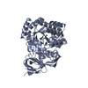
| ||||||||||
| 2 | 
| ||||||||||
| Unit cell |
| ||||||||||
| Details | The asymmetric unit contains two molecules identified as chain A and B. Each molecule is independent and biologically active. |
-Components
| #1: Protein | Mass: 50468.871 Da / Num. of mol.: 2 Source method: isolated from a genetically manipulated source Source: (gene. exp.) #2: Chemical | ChemComp-MN /   Mass: 54.938 Da / Num. of mol.: 4 / Source method: obtained synthetically / Formula: Mn Mass: 54.938 Da / Num. of mol.: 4 / Source method: obtained synthetically / Formula: Mn#3: Water | ChemComp-HOH / |  Mass: 18.015 Da / Num. of mol.: 282 / Source method: isolated from a natural source / Formula: H2O Mass: 18.015 Da / Num. of mol.: 282 / Source method: isolated from a natural source / Formula: H2O |
|---|
-Experimental details
-Experiment
| Experiment | Method: X-RAY DIFFRACTION / Number of used crystals: 1 |
|---|
- Sample preparation
| Crystal | Density Matthews: 2.43 Å3/Da / Density % sol: 48.9 % |
|---|---|
| Crystal grow | Temperature: 300 K / Method: vapor diffusion, hanging drop / pH: 8.5 Details: PEG 3350, Cesium Chloride, imidazole, pH 8.5, VAPOR DIFFUSION, HANGING DROP, temperature 300K |
-Data collection
| Diffraction | Mean temperature: 300 K |
|---|---|
| Diffraction source | Source: ROTATING ANODE / Type: RIGAKU / Wavelength: 1.5418 Å |
| Detector | Type: MARRESEARCH / Detector: IMAGE PLATE / Date: Nov 20, 2000 / Details: mirrors |
| Radiation | Protocol: SINGLE WAVELENGTH / Monochromatic (M) / Laue (L): M / Scattering type: x-ray |
| Radiation wavelength | Wavelength: 1.5418 Å / Relative weight: 1 |
| Reflection | Resolution: 2.3→50 Å / Num. all: 42440 / Num. obs: 42440 / Observed criterion σ(I): 0 |
| Reflection shell | Resolution: 2.3→2.38 Å / Rmerge(I) obs: 0.376 / Num. unique all: 4214 / % possible all: 97.3 |
- Processing
| Software |
| ||||||||||||||||||||||||||||
|---|---|---|---|---|---|---|---|---|---|---|---|---|---|---|---|---|---|---|---|---|---|---|---|---|---|---|---|---|---|
| Refinement | Method to determine structure: MOLECULAR REPLACEMENT Starting model: Modified D-hydantoinase from Thermus sp. (PDB ID 1GKP) Resolution: 2.3→50 Å / Data cutoff high absF: 10000 / Data cutoff low absF: 0 / Isotropic thermal model: overall / Cross valid method: R-free / σ(F): 0 / Stereochemistry target values: Engh & Huber
| ||||||||||||||||||||||||||||
| Displacement parameters |
| ||||||||||||||||||||||||||||
| Refinement step | Cycle: LAST / Resolution: 2.3→50 Å
| ||||||||||||||||||||||||||||
| Refine LS restraints |
|
