 Movie
Movie Controller


+ Open data

- Basic information
| Entry | Database: PDB / ID: 1yj4 | ||||||
|---|---|---|---|---|---|---|---|
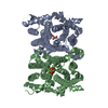
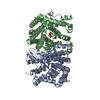
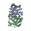
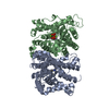
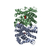
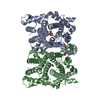
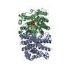
| Title | Y305F Trichodiene Synthase | ||||||
 Components Components | Trichodiene synthase | ||||||
 Keywords Keywords | LYASE / terpenoid cyclase fold / site-directed mutant | ||||||
| Function / homology |  Function and homology information Function and homology informationtrichodiene synthase / trichodiene synthase activity / sesquiterpenoid biosynthetic process / metal ion binding Similarity search - Function | ||||||
| Biological species |  Fusarium sporotrichioides (fungus) Fusarium sporotrichioides (fungus) | ||||||
| Method |  X-RAY DIFFRACTION / SYNCHROTRON / FOURIER SYNTHESIS / Resolution: 2.3 Å X-RAY DIFFRACTION / SYNCHROTRON / FOURIER SYNTHESIS / Resolution: 2.3 Å | ||||||
 Authors Authors | Vedula, L.S. / Rynkiewicz, M.J. / Pyun, H.J. / Coates, R.M. / Cane, D.E. / Christianson, D.W. | ||||||
 Citation Citation | Journal: Biochemistry / Year: 2005 Title: Molecular Recognition of the Substrate Diphosphate Group Governs Product Diversity in Trichodiene Synthase Mutants. Authors: Vedula, L.S. / Rynkiewicz, M.J. / Pyun, H.J. / Coates, R.M. / Cane, D.E. / Christianson, D.W. | ||||||
| History |
|
- Structure visualization
| Structure viewer | Molecule: MolmilJmol/JSmol |
|---|
- Downloads & links
-Download
| PDBx/mmCIF format | 1yj4.cif.gz | 154.9 KB | Display | PDBx/mmCIF format |
|---|---|---|---|---|
| PDB format | pdb1yj4.ent.gz | 124.3 KB | Display | PDB format |
| PDBx/mmJSON format | 1yj4.json.gz | Tree view | PDBx/mmJSON format | |
| Others |  Other downloads Other downloads |
-Validation report
| Arichive directory | https://data.pdbj.org/pub/pdb/validation_reports/yj/1yj4ftp://data.pdbj.org/pub/pdb/validation_reports/yj/1yj4 | HTTPS FTP |
|---|
-Related structure data
| Related structure data |  1yyqC 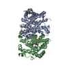 1yyrC 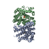 1yysC  1yytC 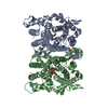 1yyuC 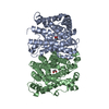 1jfaS S: Starting model for refinement C: citing same article ( |
|---|---|
| Similar structure data |


-Links
 PDBj
PDBj
- Assembly
| Deposited unit | 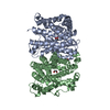
| ||||||||
|---|---|---|---|---|---|---|---|---|---|
| 1 |
| ||||||||
| Unit cell |
| ||||||||
| Details | The biological assembly is a dimer found in the asymmetric unit. |
-Components
| #1: Protein | Mass: 44034.602 Da / Num. of mol.: 2 / Mutation: Y305F Source method: isolated from a genetically manipulated source Source: (gene. exp.) Fusarium sporotrichioides (fungus) / Gene: TRI5 / Species (production host): Escherichia coli / Production host:  #2: Chemical | ChemComp-EDO /   Mass: 62.068 Da / Num. of mol.: 4 / Source method: obtained synthetically / Formula: C2H6O2 Mass: 62.068 Da / Num. of mol.: 4 / Source method: obtained synthetically / Formula: C2H6O2#3: Water | ChemComp-HOH / |  Mass: 18.015 Da / Num. of mol.: 159 / Source method: isolated from a natural source / Formula: H2O Mass: 18.015 Da / Num. of mol.: 159 / Source method: isolated from a natural source / Formula: H2O |
|---|
-Experimental details
-Experiment
| Experiment | Method: X-RAY DIFFRACTION / Number of used crystals: 1 |
|---|
- Sample preparation
| Crystal | Density Matthews: 3.72 Å3/Da / Density % sol: 66.9 % |
|---|---|
| Crystal grow | Temperature: 277 K / Method: vapor diffusion, hanging drop / pH: 6.9 Details: PEG 8000, Calcium chloride, Sodium HEPES, pH 6.9, VAPOR DIFFUSION, HANGING DROP, temperature 277K |
-Data collection
| Diffraction | Mean temperature: 100 K |
|---|---|
| Diffraction source | Source: SYNCHROTRON / Site: NSLS  / Beamline: X4A / Wavelength: 1.0721 / Wavelength: 1.0721 Å / Beamline: X4A / Wavelength: 1.0721 / Wavelength: 1.0721 Å |
| Detector | Detector: CCD / Date: Nov 14, 2003 |
| Radiation | Protocol: SINGLE WAVELENGTH / Monochromatic (M) / Laue (L): M / Scattering type: x-ray |
| Radiation wavelength | Wavelength: 1.0721 Å / Relative weight: 1 |
| Reflection | Resolution: 2.3→40 Å / Num. all: 54949 / Num. obs: 54949 / % possible obs: 93.9 % / Observed criterion σ(F): 0 / Observed criterion σ(I): 0 / Redundancy: 3 % / Biso Wilson estimate: 53.4 Å2 / Rmerge(I) obs: 0.059 / Net I/σ(I): 22.2 |
| Reflection shell | Resolution: 2.3→2.38 Å / Redundancy: 2.6 % / Rmerge(I) obs: 0.448 / Mean I/σ(I) obs: 3.1 / % possible all: 94.6 |
- Processing
| Software |
| |||||||||||||||||||||||||
|---|---|---|---|---|---|---|---|---|---|---|---|---|---|---|---|---|---|---|---|---|---|---|---|---|---|---|
| Refinement | Method to determine structure: FOURIER SYNTHESIS Starting model: PDB ID 1JFA Resolution: 2.3→40 Å / Cross valid method: THROUGHOUT / σ(F): 0 / Stereochemistry target values: Engh & Huber Details: Residues in PDB that have occupancy zero were refined as alanines.
| |||||||||||||||||||||||||
| Displacement parameters | Biso mean: 54 Å2 | |||||||||||||||||||||||||
| Refinement step | Cycle: LAST / Resolution: 2.3→40 Å
| |||||||||||||||||||||||||
| Refine LS restraints |
|
