Mass: 18.015 Da / Num. of mol.: 123 / Source method: isolated from a natural source / Formula: H2O
Has protein modification
Y
-
Experimental details
-
Experiment
Experiment
Method: X-RAY DIFFRACTION / Number of used crystals: 1
-
Sample preparation
Crystal
Density Matthews: 2.55 Å3/Da / Density % sol: 51.75 % Description: UNMODELED DENSITY APPEARS IN BOTH SUBUNITS IN THE REGION BETWEEN LYS 43 TYR 63 PROBABLY DUE TO NADP.
Crystal grow
Temperature: 277 K / Method: vapor diffusion, hanging drop / pH: 6.5 Details: 10 MG/ML PROTEIN, 15% PEG4K, 0.168 M SODIUM CHLORIDE, 0.050 M PIPES, VAPOR DIFFUSION, HANGING DROP, temperature 277K, pH 6.5
Method to determine structure: SAD / Resolution: 2.488→31.06 Å / Cor.coef. Fo:Fc: 0.927 / Cor.coef. Fo:Fc free: 0.873 / WRfactor Rfree: 0.283 / WRfactor Rwork: 0.218 / SU B: 10.133 / SU ML: 0.229 / SU R Cruickshank DPI: 0.552 / Cross valid method: THROUGHOUT / ESU R: 0.552 / ESU R Free: 0.321 / Stereochemistry target values: MAXIMUM LIKELIHOOD Details: HYDROGENS HAVE BEEN ADDED IN THE RIDING POSITIONS, SELENIUM C COEFFICIENT FOR STRUCTURE FACTOR CALCULATION SET TO -9.0000, MOLPROBITY USED TO ASSIST IN FINAL MODEL BUILDING
Rfactor
Num. reflection
% reflection
Selection details
Rfree
0.28254
1472
5.1 %
RANDOM
Rwork
0.21822
-
-
-
all
0.22155
-
-
-
obs
-
27390
98.351 %
-
Solvent computation
Ion probe radii: 0.8 Å / Shrinkage radii: 0.8 Å / VDW probe radii: 1.2 Å / Solvent model: MASK BULK SOLVENT
Displacement parameters
Biso mean: 33.072 Å2
Baniso -1
Baniso -2
Baniso -3
1-
1.389 Å2
0 Å2
0 Å2
2-
-
2.068 Å2
0 Å2
3-
-
-
-3.457 Å2
Refinement step
Cycle: LAST / Resolution: 2.488→31.06 Å
Protein
Nucleic acid
Ligand
Solvent
Total
Num. atoms
5340
0
0
123
5463
Refine LS restraints
Refine-ID
Type
Dev ideal
Dev ideal target
Number
X-RAY DIFFRACTION
r_bond_refined_d
0.022
0.022
5453
X-RAY DIFFRACTION
r_angle_refined_deg
2.05
1.954
7408
X-RAY DIFFRACTION
r_dihedral_angle_1_deg
8.041
5
689
X-RAY DIFFRACTION
r_dihedral_angle_2_deg
40.318
24.554
224
X-RAY DIFFRACTION
r_dihedral_angle_3_deg
19.93
15
919
X-RAY DIFFRACTION
r_dihedral_angle_4_deg
21.043
15
25
X-RAY DIFFRACTION
r_chiral_restr
0.162
0.2
854
X-RAY DIFFRACTION
r_gen_planes_refined
0.008
0.02
4061
X-RAY DIFFRACTION
r_nbd_refined
0.277
0.2
2846
X-RAY DIFFRACTION
r_nbtor_refined
0.326
0.2
3753
X-RAY DIFFRACTION
r_xyhbond_nbd_refined
0.216
0.2
289
X-RAY DIFFRACTION
r_symmetry_vdw_refined
0.238
0.2
14
X-RAY DIFFRACTION
r_symmetry_hbond_refined
0.34
0.2
3
X-RAY DIFFRACTION
r_mcbond_it
1.757
2
3550
X-RAY DIFFRACTION
r_mcangle_it
3.216
4
5566
X-RAY DIFFRACTION
r_scbond_it
5.826
6
2195
X-RAY DIFFRACTION
r_scangle_it
7.878
8
1842
LS refinement shell
Refine-ID: X-RAY DIFFRACTION / Total num. of bins used: 20
Resolution (Å)
Rfactor Rfree
Num. reflection Rfree
Rfactor Rwork
Num. reflection Rwork
Num. reflection all
% reflection obs (%)
2.488-2.553
0.374
86
0.285
1651
2127
81.664
2.553-2.622
0.407
101
0.255
1915
2075
97.157
2.622-2.697
0.327
95
0.244
1889
2003
99.051
2.697-2.78
0.352
106
0.266
1870
1979
99.848
2.78-2.87
0.324
100
0.244
1772
1873
99.947
2.87-2.97
0.317
87
0.233
1756
1845
99.892
2.97-3.081
0.303
84
0.226
1706
1790
100
3.081-3.206
0.314
87
0.217
1620
1707
100
3.206-3.347
0.245
76
0.215
1572
1648
100
3.347-3.508
0.259
81
0.209
1512
1593
100
3.508-3.695
0.265
76
0.213
1435
1511
100
3.695-3.916
0.252
79
0.202
1353
1432
100
3.916-4.181
0.226
95
0.191
1271
1366
100
4.181-4.509
0.233
54
0.189
1200
1254
100
4.509-4.93
0.224
51
0.184
1131
1183
99.915
4.93-5.494
0.273
64
0.207
996
1060
100
5.494-6.311
0.315
48
0.224
914
962
100
6.311-7.651
0.28
44
0.232
792
836
100
7.651-10.506
0.245
36
0.168
623
660
99.848
10.506-31.06
0.424
22
0.344
412
442
98.19
+
About Yorodumi
-
News
-
Feb 9, 2022. New format data for meta-information of EMDB entries
New format data for meta-information of EMDB entries
Version 3 of the EMDB header file is now the official format.
The previous official version 1.9 will be removed from the archive.
In the structure databanks used in Yorodumi, some data are registered as the other names, "COVID-19 virus" and "2019-nCoV". Here are the details of the virus and the list of structure data.
Jan 31, 2019. EMDB accession codes are about to change! (news from PDBe EMDB page)
EMDB accession codes are about to change! (news from PDBe EMDB page)
The allocation of 4 digits for EMDB accession codes will soon come to an end. Whilst these codes will remain in use, new EMDB accession codes will include an additional digit and will expand incrementally as the available range of codes is exhausted. The current 4-digit format prefixed with “EMD-” (i.e. EMD-XXXX) will advance to a 5-digit format (i.e. EMD-XXXXX), and so on. It is currently estimated that the 4-digit codes will be depleted around Spring 2019, at which point the 5-digit format will come into force.
The EM Navigator/Yorodumi systems omit the EMD- prefix.
Related info.:Q: What is EMD? / ID/Accession-code notation in Yorodumi/EM Navigator
Yorodumi is a browser for structure data from EMDB, PDB, SASBDB, etc.
This page is also the successor to EM Navigator detail page, and also detail information page/front-end page for Omokage search.
The word "yorodu" (or yorozu) is an old Japanese word meaning "ten thousand". "mi" (miru) is to see.
Related info.:EMDB / PDB / SASBDB / Comparison of 3 databanks / Yorodumi Search / Aug 31, 2016. New EM Navigator & Yorodumi / Yorodumi Papers / Jmol/JSmol / Function and homology information / Changes in new EM Navigator and Yorodumi
 Movie
Movie Controller

 Yorodumi
 Open data
 Basic information
 Components
 Keywords
 Function and homology information
 X-RAY DIFFRACTION /
  Authors
 Citation
 Structure visualization
 Downloads & links
 Other downloads

 PDBj
PDBj Assembly
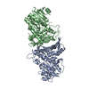
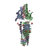
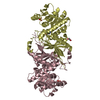
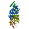
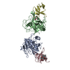
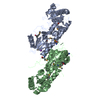
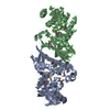

 Mass: 18.015 Da / Num. of mol.: 123 / Source method: isolated from a natural source / Formula: H2O
 Sample preparation
 / Beamline: 14-ID-B / Wavelength: 0.97927 Å
 Processing