 Movie
Movie Controller

[English] 日本語
 Yorodumi
- PDB-1yaa: ASPARTATE AMINOTRANSFERASE FROM SACCHAROMYCES CEREVISIAE CYTOPLASM -
+ Open data

- Basic information
| Entry | Database: PDB / ID: 1yaa | ||||||
|---|---|---|---|---|---|---|---|
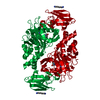
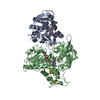
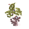
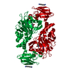
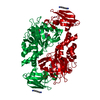
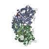
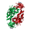
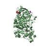
| Title | ASPARTATE AMINOTRANSFERASE FROM SACCHAROMYCES CEREVISIAE CYTOPLASM | ||||||
 Components Components | ASPARTATE AMINOTRANSFERASE | ||||||
 Keywords Keywords | AMINOTRANSFERASE / TRANSFERASE | ||||||
| Function / homology |  Function and homology information Function and homology informationAspartate and asparagine metabolism / Malate-aspartate shuttle / negative regulation of translation in response to oxidative stress / L-aspartate biosynthetic process / aspartate metabolic process / glutamate metabolic process / aspartate transaminase / L-aspartate:2-oxoglutarate transaminase activity / 2-oxoglutarate metabolic process / ribosomal large subunit binding ...Aspartate and asparagine metabolism / Malate-aspartate shuttle / negative regulation of translation in response to oxidative stress / L-aspartate biosynthetic process / aspartate metabolic process / glutamate metabolic process / aspartate transaminase / L-aspartate:2-oxoglutarate transaminase activity / 2-oxoglutarate metabolic process / ribosomal large subunit binding / pyridoxal phosphate binding / peroxisome / cytosol Similarity search - Function | ||||||
| Biological species |  | ||||||
| Method |  X-RAY DIFFRACTION / MOLECULAR REPLACEMENT / Resolution: 2.05 Å X-RAY DIFFRACTION / MOLECULAR REPLACEMENT / Resolution: 2.05 Å | ||||||
 Authors Authors | Jeffery, C.J. | ||||||
 Citation Citation | Journal: Protein Sci. / Year: 1998 Title: Crystal structure of Saccharomyces cerevisiae cytosolic aspartate aminotransferase. Authors: Jeffery, C.J. / Barry, T. / Doonan, S. / Petsko, G.A. / Ringe, D. #1: Journal: To be PublishedTitle: Crystallization and Preliminary X-Ray Diffraction Analysis of Aspartate Aminotransferase from Saccharomyces Cerevisiae Authors: Jeffery, C.J. / Barry, T. / Doonan, S. / Petsko, G.A. / Ringe, D. | ||||||
| History |
|
- Structure visualization
| Structure viewer | Molecule: MolmilJmol/JSmol |
|---|
- Downloads & links
-Download
| PDBx/mmCIF format | 1yaa.cif.gz | 329.6 KB | Display | PDBx/mmCIF format |
|---|---|---|---|---|
| PDB format | pdb1yaa.ent.gz | 271.2 KB | Display | PDB format |
| PDBx/mmJSON format | 1yaa.json.gz | Tree view | PDBx/mmJSON format | |
| Others |  Other downloads Other downloads |
-Validation report
| Arichive directory | https://data.pdbj.org/pub/pdb/validation_reports/ya/1yaaftp://data.pdbj.org/pub/pdb/validation_reports/ya/1yaa | HTTPS FTP |
|---|
-Related structure data
| Related structure data |  2cstS S: Starting model for refinement |
|---|---|
| Similar structure data |


-Links
 PDBj
PDBj- Assembly
| Deposited unit | 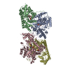
| ||||||||
|---|---|---|---|---|---|---|---|---|---|
| 1 | 
| ||||||||
| 2 | 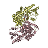
| ||||||||
| Unit cell |
|
-Components
| #1: Protein | Mass: 45361.547 Da / Num. of mol.: 4 / Source method: isolated from a natural source Details: PYRIDOXAL PHOSPHATE COFACTOR COVALENTLY BOUND TO LYS 258 VIA SCHIFF BASE LINKAGE IN EACH SUBUNIT Source: (natural) #2: Chemical | ChemComp-MAE /   Mass: 116.072 Da / Num. of mol.: 4 / Source method: obtained synthetically / Formula: C4H4O4 Mass: 116.072 Da / Num. of mol.: 4 / Source method: obtained synthetically / Formula: C4H4O4#3: Chemical | ChemComp-PLP /   Mass: 247.142 Da / Num. of mol.: 4 / Source method: obtained synthetically / Formula: C8H10NO6P Mass: 247.142 Da / Num. of mol.: 4 / Source method: obtained synthetically / Formula: C8H10NO6P#4: Water | ChemComp-HOH / |  Mass: 18.015 Da / Num. of mol.: 716 / Source method: isolated from a natural source / Formula: H2O Mass: 18.015 Da / Num. of mol.: 716 / Source method: isolated from a natural source / Formula: H2O |
|---|
-Experimental details
-Experiment
| Experiment | Method: X-RAY DIFFRACTION / Number of used crystals: 1 |
|---|
- Sample preparation
| Crystal | Density Matthews: 2.4 Å3/Da / Density % sol: 49 % | |||||||||||||||||||||||||||||||||||||||||||||
|---|---|---|---|---|---|---|---|---|---|---|---|---|---|---|---|---|---|---|---|---|---|---|---|---|---|---|---|---|---|---|---|---|---|---|---|---|---|---|---|---|---|---|---|---|---|---|
| Crystal grow | pH: 4.6 Details: PROTEIN SOLUTION CONTAINED 5-15MG PROTEIN, 10MM PMSF, 10MM PLP, 10MM SODIUM ACETATE BUFFER, PH 5.8, AND 100MM MALEATE. RESERVOIR SOLUTION CONTAINED 0.2 M AMMONIUM ACETATE, 0.1 M SODIUM ...Details: PROTEIN SOLUTION CONTAINED 5-15MG PROTEIN, 10MM PMSF, 10MM PLP, 10MM SODIUM ACETATE BUFFER, PH 5.8, AND 100MM MALEATE. RESERVOIR SOLUTION CONTAINED 0.2 M AMMONIUM ACETATE, 0.1 M SODIUM ACETATE BUFFER, PH 4.6, AND 20-22% PEG 4000. PH range: 4.6-5.8 | |||||||||||||||||||||||||||||||||||||||||||||
| Crystal | *PLUS | |||||||||||||||||||||||||||||||||||||||||||||
| Crystal grow | *PLUS pH: 5.8 / Method: vapor diffusion, hanging drop / Details: Jeffery, C.J., (1998) Acta Cryst., D54, 659. | |||||||||||||||||||||||||||||||||||||||||||||
| Components of the solutions | *PLUS
|
-Data collection
| Diffraction | Mean temperature: 297 K |
|---|---|
| Diffraction source | Source: ROTATING ANODE / Type: RIGAKU RUH2R / Wavelength: 1.5418 |
| Detector | Type: RIGAKU / Detector: IMAGE PLATE / Date: Oct 1, 1992 |
| Radiation | Monochromator: NI FILTER / Monochromatic (M) / Laue (L): M / Scattering type: x-ray |
| Radiation wavelength | Wavelength: 1.5418 Å / Relative weight: 1 |
| Reflection | Highest resolution: 2.05 Å / Num. obs: 470081 / % possible obs: 93 % / Observed criterion σ(I): 0 / Rmerge(I) obs: 0.12 / Rsym value: 0.12 |
| Reflection shell | Resolution: 2.05→2.14 Å / % possible all: 87 |
| Reflection | *PLUS Num. obs: 100347 / % possible obs: 93 % / Num. measured all: 470081 |
| Reflection shell | *PLUS % possible obs: 88 % |
- Processing
| Software |
| ||||||||||||||||||||||||||||||||||||||||||||||||||||||||||||
|---|---|---|---|---|---|---|---|---|---|---|---|---|---|---|---|---|---|---|---|---|---|---|---|---|---|---|---|---|---|---|---|---|---|---|---|---|---|---|---|---|---|---|---|---|---|---|---|---|---|---|---|---|---|---|---|---|---|---|---|---|---|
| Refinement | Method to determine structure: MOLECULAR REPLACEMENT Starting model: PDB ENTRY 2CST Resolution: 2.05→10 Å / Rfactor Rfree error: 0.003 / Data cutoff high absF: 1000000 / Data cutoff low absF: 0 / Cross valid method: THROUGHOUT / σ(F): 0
| ||||||||||||||||||||||||||||||||||||||||||||||||||||||||||||
| Displacement parameters | Biso mean: 19.9 Å2 | ||||||||||||||||||||||||||||||||||||||||||||||||||||||||||||
| Refinement step | Cycle: LAST / Resolution: 2.05→10 Å
| ||||||||||||||||||||||||||||||||||||||||||||||||||||||||||||
| Refine LS restraints |
| ||||||||||||||||||||||||||||||||||||||||||||||||||||||||||||
| LS refinement shell | Resolution: 2.05→2.14 Å / Rfactor Rfree error: 0.001 / Total num. of bins used: 8
| ||||||||||||||||||||||||||||||||||||||||||||||||||||||||||||
| Xplor file |
| ||||||||||||||||||||||||||||||||||||||||||||||||||||||||||||
| Software | *PLUS Name: X-PLOR / Version: 3.851 / Classification: refinement | ||||||||||||||||||||||||||||||||||||||||||||||||||||||||||||
| Refine LS restraints | *PLUS
|
