 Movie
Movie Controller

[English] 日本語
 Yorodumi
- PDB-4bgk: Three dimensional structure of human gamma-butyrobetaine hydroxyl... -
+ Open data

- Basic information
| Entry | Database: PDB / ID: 4bgk | ||||||
|---|---|---|---|---|---|---|---|
| Title | Three dimensional structure of human gamma-butyrobetaine hydroxylase in complex with (3-(Trimethylammonio)propyl)phosphinate | ||||||
 Components Components | GAMMA-BUTYROBETAINE DIOXYGENASE | ||||||
 Keywords Keywords | OXIDOREDUCTASE | ||||||
| Function / homology |  Function and homology information Function and homology informationgamma-butyrobetaine dioxygenase / gamma-butyrobetaine dioxygenase activity / Carnitine synthesis / carnitine biosynthetic process / iron ion binding / mitochondrion / extracellular exosome / zinc ion binding / identical protein binding / cytosol Similarity search - Function | ||||||
| Biological species |  HOMO SAPIENS (human) HOMO SAPIENS (human) | ||||||
| Method |  X-RAY DIFFRACTION / SYNCHROTRON / MOLECULAR REPLACEMENT / Resolution: 2.18 Å X-RAY DIFFRACTION / SYNCHROTRON / MOLECULAR REPLACEMENT / Resolution: 2.18 Å | ||||||
 Authors Authors | Tars, K. / Leitans, J. / Kazaks, A. | ||||||
 Citation Citation | Journal: J.Med.Chem. / Year: 2014 Title: Targeting Carnitine Biosynthesis: Discovery of New Inhibitors Against Gamma-Butyrobetaine Hydroxylase. Authors: Tars, K. / Leitans, J. / Kazaks, A. / Zelencova, D. / Liepinsh, E. / Kuka, J. / Makrecka, M. / Lola, D. / Andrianovs, V. / Gustina, D. / Grinberga, S. / Liepinsh, E. / Kalvinsh, I. / ...Authors: Tars, K. / Leitans, J. / Kazaks, A. / Zelencova, D. / Liepinsh, E. / Kuka, J. / Makrecka, M. / Lola, D. / Andrianovs, V. / Gustina, D. / Grinberga, S. / Liepinsh, E. / Kalvinsh, I. / Dambrova, M. / Loza, E. / Pugovics, O. | ||||||
| History |
|
- Structure visualization
| Structure viewer | Molecule: MolmilJmol/JSmol |
|---|
- Downloads & links
-Download
| PDBx/mmCIF format | 4bgk.cif.gz | 103.2 KB | Display | PDBx/mmCIF format |
|---|---|---|---|---|
| PDB format | pdb4bgk.ent.gz | 77 KB | Display | PDB format |
| PDBx/mmJSON format | 4bgk.json.gz | Tree view | PDBx/mmJSON format | |
| Others |  Other downloads Other downloads |
-Validation report
| Arichive directory | https://data.pdbj.org/pub/pdb/validation_reports/bg/4bgkftp://data.pdbj.org/pub/pdb/validation_reports/bg/4bgk | HTTPS FTP |
|---|
-Related structure data
| Related structure data |  4bg1C  4bgmC  4bhfC  4bhiC  4c5wC  3o2gS C: citing same article ( S: Starting model for refinement |
|---|---|
| Similar structure data |


-Links
 PDBj
PDBj
- Assembly
| Deposited unit | 
| ||||||||
|---|---|---|---|---|---|---|---|---|---|
| 1 | 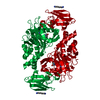
| ||||||||
| Unit cell |
| ||||||||
| Components on special symmetry positions |
|
-Components
-Protein , 1 types, 1 molecules A
| #1: Protein | Mass: 44774.867 Da / Num. of mol.: 1 Source method: isolated from a genetically manipulated source Source: (gene. exp.) HOMO SAPIENS (human) / Production host:  References: UniProt: O75936, gamma-butyrobetaine dioxygenase |
|---|
-Non-polymers , 5 types, 373 molecules 


| #2: Chemical | ChemComp-OGA /  Mass: 147.086 Da / Num. of mol.: 1 / Source method: obtained synthetically / Formula: C4H5NO5 Mass: 147.086 Da / Num. of mol.: 1 / Source method: obtained synthetically / Formula: C4H5NO5 | ||||
|---|---|---|---|---|---|
| #3: Chemical | ChemComp-RTK /  Mass: 166.178 Da / Num. of mol.: 1 / Source method: obtained synthetically / Formula: C6H17NO2P Mass: 166.178 Da / Num. of mol.: 1 / Source method: obtained synthetically / Formula: C6H17NO2P | ||||
| #4: Chemical |  Mass: 65.409 Da / Num. of mol.: 2 / Source method: obtained synthetically / Formula: Zn Mass: 65.409 Da / Num. of mol.: 2 / Source method: obtained synthetically / Formula: Zn#5: Chemical | ChemComp-16D / |  Mass: 116.205 Da / Num. of mol.: 1 / Source method: obtained synthetically / Formula: C6H16N2 Mass: 116.205 Da / Num. of mol.: 1 / Source method: obtained synthetically / Formula: C6H16N2#6: Water | ChemComp-HOH / | Mass: 18.015 Da / Num. of mol.: 368 / Source method: isolated from a natural source / Formula: H2O |
-Experimental details
-Experiment
| Experiment | Method: X-RAY DIFFRACTION / Number of used crystals: 1 |
|---|
- Sample preparation
| Crystal | Density Matthews: 2.5 Å3/Da / Density % sol: 52 % / Description: NONE |
|---|---|
| Crystal grow | Temperature: 294 K / Method: vapor diffusion, sitting drop / pH: 7 Details: 20 % PEG 3350, 1.5 % 1.6-DIAMINOHEXANE, 200 MM AMMONIUM CITRATE PH 7.0, 10 MM ZNSO4, 8 MM N-OXALYLGLYCINE, 4 MM INHIBITOR, PROTEIN 6.5 MG/ML, VAPOR DIFFUSION, SITTING DROP, TEMPERATURE 294K, TIME 2-7 DAYS |
-Data collection
| Diffraction | Mean temperature: 100 K |
|---|---|
| Diffraction source | Source: SYNCHROTRON / Site: MAX II  / Beamline: I911-3 / Wavelength: 1.01 / Beamline: I911-3 / Wavelength: 1.01 |
| Detector | Type: MARRESEARCH / Detector: CCD / Date: Jun 5, 2011 / Details: MIRRORS |
| Radiation | Monochromator: DOUBLE SI CRYSTAL MONOCHROMATOR / Protocol: SINGLE WAVELENGTH / Monochromatic (M) / Laue (L): M / Scattering type: x-ray |
| Radiation wavelength | Wavelength: 1.01 Å / Relative weight: 1 |
| Reflection | Resolution: 2.18→37.55 Å / Num. obs: 23613 / % possible obs: 97.9 % / Redundancy: 2.5 % / Biso Wilson estimate: 21.614 Å2 / Rmerge(I) obs: 0.13 / Net I/σ(I): 6.2 |
| Reflection shell | Resolution: 2.18→2.3 Å / Redundancy: 2.5 % / Rmerge(I) obs: 0.45 / Mean I/σ(I) obs: 2.2 / % possible all: 99.5 |
- Processing
| Software |
| ||||||||||||||||||||||||||||||||||||||||||||||||||||||||||||||||||||||||||||||||||||||||||||||||||||||||||||||||||||||||||||||||||||||||||||||||||||||||||||||||||||||||||||||||||||||
|---|---|---|---|---|---|---|---|---|---|---|---|---|---|---|---|---|---|---|---|---|---|---|---|---|---|---|---|---|---|---|---|---|---|---|---|---|---|---|---|---|---|---|---|---|---|---|---|---|---|---|---|---|---|---|---|---|---|---|---|---|---|---|---|---|---|---|---|---|---|---|---|---|---|---|---|---|---|---|---|---|---|---|---|---|---|---|---|---|---|---|---|---|---|---|---|---|---|---|---|---|---|---|---|---|---|---|---|---|---|---|---|---|---|---|---|---|---|---|---|---|---|---|---|---|---|---|---|---|---|---|---|---|---|---|---|---|---|---|---|---|---|---|---|---|---|---|---|---|---|---|---|---|---|---|---|---|---|---|---|---|---|---|---|---|---|---|---|---|---|---|---|---|---|---|---|---|---|---|---|---|---|---|---|
| Refinement | Method to determine structure: MOLECULAR REPLACEMENT Starting model: PDB ENTRY 3O2G Resolution: 2.18→42.44 Å / Cor.coef. Fo:Fc: 0.962 / Cor.coef. Fo:Fc free: 0.934 / SU B: 4.173 / SU ML: 0.107 / Cross valid method: THROUGHOUT / ESU R: 0.21 / ESU R Free: 0.171 / Stereochemistry target values: MAXIMUM LIKELIHOOD / Details: HYDROGENS HAVE BEEN ADDED IN THE RIDING POSITIONS.
| ||||||||||||||||||||||||||||||||||||||||||||||||||||||||||||||||||||||||||||||||||||||||||||||||||||||||||||||||||||||||||||||||||||||||||||||||||||||||||||||||||||||||||||||||||||||
| Solvent computation | Ion probe radii: 0.8 Å / Shrinkage radii: 0.8 Å / VDW probe radii: 1.2 Å / Solvent model: MASK | ||||||||||||||||||||||||||||||||||||||||||||||||||||||||||||||||||||||||||||||||||||||||||||||||||||||||||||||||||||||||||||||||||||||||||||||||||||||||||||||||||||||||||||||||||||||
| Displacement parameters | Biso mean: 21.266 Å2
| ||||||||||||||||||||||||||||||||||||||||||||||||||||||||||||||||||||||||||||||||||||||||||||||||||||||||||||||||||||||||||||||||||||||||||||||||||||||||||||||||||||||||||||||||||||||
| Refinement step | Cycle: LAST / Resolution: 2.18→42.44 Å
| ||||||||||||||||||||||||||||||||||||||||||||||||||||||||||||||||||||||||||||||||||||||||||||||||||||||||||||||||||||||||||||||||||||||||||||||||||||||||||||||||||||||||||||||||||||||
| Refine LS restraints |
|