Method to determine structure: MIR / Resolution: 1.78→28.98 Å / Cor.coef. Fo:Fc: 0.969 / Cor.coef. Fo:Fc free: 0.955 / SU B: 3.583 / SU ML: 0.062 / Cross valid method: THROUGHOUT / ESU R Free: 0.095 / Stereochemistry target values: MAXIMUM LIKELIHOOD / Details: HYDROGENS HAVE BEEN USED IF PRESENT IN THE INPUT
Rfactor
Num. reflection
% reflection
Selection details
Rfree
0.17633
2226
5.1 %
RANDOM
Rwork
0.14717
-
-
-
all
0.14869
43809
-
-
obs
0.14869
41560
99.91 %
-
Solvent computation
Ion probe radii: 0.8 Å / Shrinkage radii: 0.8 Å / VDW probe radii: 1.2 Å / Solvent model: MASK
Displacement parameters
Biso mean: 16.971 Å2
Baniso -1
Baniso -2
Baniso -3
1-
0.29 Å2
0.15 Å2
0 Å2
2-
-
0.29 Å2
0 Å2
3-
-
-
-0.44 Å2
Refinement step
Cycle: LAST / Resolution: 1.78→28.98 Å
Protein
Nucleic acid
Ligand
Solvent
Total
Num. atoms
3109
0
42
437
3588
Refine LS restraints
Refine-ID
Type
Dev ideal
Dev ideal target
Number
X-RAY DIFFRACTION
r_bond_refined_d
0.016
0.022
3321
X-RAY DIFFRACTION
r_bond_other_d
0.001
0.02
2260
X-RAY DIFFRACTION
r_angle_refined_deg
1.554
1.95
4519
X-RAY DIFFRACTION
r_angle_other_deg
0.926
3
5507
X-RAY DIFFRACTION
r_dihedral_angle_1_deg
6.356
5
418
X-RAY DIFFRACTION
r_dihedral_angle_2_deg
34.982
24.074
162
X-RAY DIFFRACTION
r_dihedral_angle_3_deg
12.366
15
566
X-RAY DIFFRACTION
r_dihedral_angle_4_deg
19.71
15
19
X-RAY DIFFRACTION
r_chiral_restr
0.098
0.2
491
X-RAY DIFFRACTION
r_gen_planes_refined
0.008
0.021
3714
X-RAY DIFFRACTION
r_gen_planes_other
0.001
0.02
704
LS refinement shell
Resolution: 1.78→1.826 Å / Total num. of bins used: 20
Rfactor
Num. reflection
% reflection
Rfree
0.297
179
-
Rwork
0.267
3018
-
obs
-
-
100 %
Refinement TLS params.
Method: refined / Refine-ID: X-RAY DIFFRACTION
ID
L11 (°2)
L12 (°2)
L13 (°2)
L22 (°2)
L23 (°2)
L33 (°2)
S11 (Å °)
S12 (Å °)
S13 (Å °)
S21 (Å °)
S22 (Å °)
S23 (Å °)
S31 (Å °)
S32 (Å °)
S33 (Å °)
T11 (Å2)
T12 (Å2)
T13 (Å2)
T22 (Å2)
T23 (Å2)
T33 (Å2)
Origin x (Å)
Origin y (Å)
Origin z (Å)
1
1.8846
0.0805
-0.8519
0.6538
0.3784
2.0847
0.0019
0.0866
0.0335
-0.0321
-0.0628
0.2041
-0.1005
-0.2627
0.0609
0.0319
0.0235
0.0065
0.0412
-0.0112
0.0771
-10.4397
29.8216
123.8685
2
0.7789
-0.2867
-0.0481
1.0538
0.0807
0.7927
-0.0444
0.0496
0.0052
-0.0272
0.0213
0.085
-0.1232
-0.0565
0.0231
0.0269
0.0152
-0.0068
0.0477
0.0035
0.0084
12.4256
30.2127
87.7316
Refinement TLS group
ID
Refine-ID
Refine TLS-ID
Auth asym-ID
Auth seq-ID
1
X-RAY DIFFRACTION
1
A
1 - 108
2
X-RAY DIFFRACTION
2
A
109 - 384
+
About Yorodumi
-
News
-
Feb 9, 2022. New format data for meta-information of EMDB entries
New format data for meta-information of EMDB entries
Version 3 of the EMDB header file is now the official format.
The previous official version 1.9 will be removed from the archive.
In the structure databanks used in Yorodumi, some data are registered as the other names, "COVID-19 virus" and "2019-nCoV". Here are the details of the virus and the list of structure data.
Jan 31, 2019. EMDB accession codes are about to change! (news from PDBe EMDB page)
EMDB accession codes are about to change! (news from PDBe EMDB page)
The allocation of 4 digits for EMDB accession codes will soon come to an end. Whilst these codes will remain in use, new EMDB accession codes will include an additional digit and will expand incrementally as the available range of codes is exhausted. The current 4-digit format prefixed with “EMD-” (i.e. EMD-XXXX) will advance to a 5-digit format (i.e. EMD-XXXXX), and so on. It is currently estimated that the 4-digit codes will be depleted around Spring 2019, at which point the 5-digit format will come into force.
The EM Navigator/Yorodumi systems omit the EMD- prefix.
Related info.:Q: What is EMD? / ID/Accession-code notation in Yorodumi/EM Navigator
Yorodumi is a browser for structure data from EMDB, PDB, SASBDB, etc.
This page is also the successor to EM Navigator detail page, and also detail information page/front-end page for Omokage search.
The word "yorodu" (or yorozu) is an old Japanese word meaning "ten thousand". "mi" (miru) is to see.
Related info.:EMDB / PDB / SASBDB / Comparison of 3 databanks / Yorodumi Search / Aug 31, 2016. New EM Navigator & Yorodumi / Yorodumi Papers / Jmol/JSmol / Function and homology information / Changes in new EM Navigator and Yorodumi
 Movie
Movie Controller

 Yorodumi
 Open data
 Basic information
 Components
 Keywords
 Function and homology information
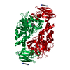
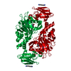
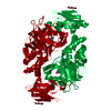
 Homo sapiens (human)
 X-RAY DIFFRACTION /
  Authors
 Citation
 Structure visualization
 Downloads & links
 Other downloads
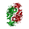


 PDBj
PDBj
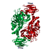
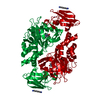
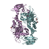
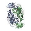
 Assembly
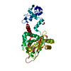
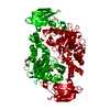
 TRICHOPLUSIA NI (cabbage looper) / Strain (production host): DH10BAC


 Mass: 65.409 Da / Num. of mol.: 2 / Source method: obtained synthetically / Formula: Zn
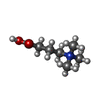
 Mass: 147.086 Da / Num. of mol.: 1 / Source method: obtained synthetically / Formula: C4H5NO5 / Comment: inhibitor*YM
 Mass: 146.207 Da / Num. of mol.: 1 / Source method: obtained synthetically / Formula: C7H16NO2
 Mass: 62.068 Da / Num. of mol.: 5 / Source method: obtained synthetically / Formula: C2H6O2
 Sample preparation
 / Beamline: I02
 Processing