 Movie
Movie Controller

[English] 日本語
 Yorodumi
- PDB-1y9w: Structural Genomics, 1.9A crystal structure of an acetyltransfera... -
+ Open data

- Basic information
| Entry | Database: PDB / ID: 1y9w | ||||||
|---|---|---|---|---|---|---|---|
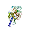
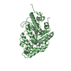
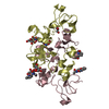
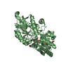
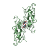
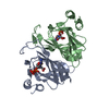
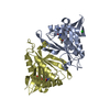
| Title | Structural Genomics, 1.9A crystal structure of an acetyltransferase from Bacillus cereus ATCC 14579 | ||||||
 Components Components | Acetyltransferase | ||||||
 Keywords Keywords | TRANSFERASE / Bacillus cereus / acetyltransferase / Structural Genomics / Protein Structure Initiative / PSI / Midwest Center for Structural Genomics / MCSG | ||||||
| Function / homology |  Function and homology information Function and homology informationacyltransferase activity, transferring groups other than amino-acyl groups / Transferases; Acyltransferases; Transferring groups other than aminoacyl groups Similarity search - Function | ||||||
| Biological species |  | ||||||
| Method |  X-RAY DIFFRACTION / SYNCHROTRON / MAD / Resolution: 1.9 Å X-RAY DIFFRACTION / SYNCHROTRON / MAD / Resolution: 1.9 Å | ||||||
 Authors Authors | Zhang, R. / Li, H. / Collart, F. / Joachimiak, A. / Midwest Center for Structural Genomics (MCSG) | ||||||
 Citation Citation | Journal: To be Published / Year: 2005 Title: 1.9A crystal structure of an acetyltransferase from Bacillus cereus ATCC 14579 Authors: Zhang, R. / Li, H. / Collart, F. / Joachimiak, A. | ||||||
| History |
|
- Structure visualization
| Structure viewer | Molecule: MolmilJmol/JSmol |
|---|
- Downloads & links
-Download
| PDBx/mmCIF format | 1y9w.cif.gz | 73.3 KB | Display | PDBx/mmCIF format |
|---|---|---|---|---|
| PDB format | pdb1y9w.ent.gz | 55.1 KB | Display | PDB format |
| PDBx/mmJSON format | 1y9w.json.gz | Tree view | PDBx/mmJSON format | |
| Others |  Other downloads Other downloads |
-Validation report
| Arichive directory | https://data.pdbj.org/pub/pdb/validation_reports/y9/1y9wftp://data.pdbj.org/pub/pdb/validation_reports/y9/1y9w | HTTPS FTP |
|---|
-Related structure data
| Similar structure data | |
|---|---|
| Other databases |


-Links
 PDBj
PDBj

- Assembly
| Deposited unit | 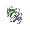
| ||||||||||
|---|---|---|---|---|---|---|---|---|---|---|---|
| 1 |
| ||||||||||
| Unit cell |
| ||||||||||
| Details | This protein existed as dimer, the Mol.A and Mol.B represent the dimer in the assymmetric unit. |
-Components
| #1: Protein | Mass: 16485.754 Da / Num. of mol.: 2 Source method: isolated from a genetically manipulated source Source: (gene. exp.) References: UniProt: Q81CG1, Transferases; Acyltransferases; Transferring groups other than aminoacyl groups #2: Water | ChemComp-HOH / |  Mass: 18.015 Da / Num. of mol.: 250 / Source method: isolated from a natural source / Formula: H2O Mass: 18.015 Da / Num. of mol.: 250 / Source method: isolated from a natural source / Formula: H2O |
|---|
-Experimental details
-Experiment
| Experiment | Method: X-RAY DIFFRACTION / Number of used crystals: 1 |
|---|
- Sample preparation
| Crystal | Density Matthews: 2.506 Å3/Da / Density % sol: 49 % |
|---|---|
| Crystal grow | Temperature: 298 K / Method: vapor diffusion, sitting drop / pH: 7.5 Details: 0.05M MgCl2, 0.1M HEPES, 30% PEG MME550, pH 7.5, VAPOR DIFFUSION, SITTING DROP, temperature 298K |
-Data collection
| Diffraction | Mean temperature: 100 K | ||||||||||||
|---|---|---|---|---|---|---|---|---|---|---|---|---|---|
| Diffraction source | Source: SYNCHROTRON / Site: APS  / Beamline: 19-ID / Wavelength: 0.9795,0.9797,0.94656 / Beamline: 19-ID / Wavelength: 0.9795,0.9797,0.94656 | ||||||||||||
| Detector | Type: SBC-2 / Detector: CCD / Date: Feb 18, 2004 / Details: mirrors | ||||||||||||
| Radiation | Monochromator: Si 111 channel / Protocol: MAD / Monochromatic (M) / Laue (L): M / Scattering type: x-ray | ||||||||||||
| Radiation wavelength |
| ||||||||||||
| Reflection | Resolution: 1.9→40 Å / Num. all: 49031 / Num. obs: 45795 / % possible obs: 93.4 % / Observed criterion σ(F): 2 / Observed criterion σ(I): 2 / Redundancy: 4.5 % / Biso Wilson estimate: 12.2 Å2 / Rmerge(I) obs: 0.074 / Net I/σ(I): 30.4 | ||||||||||||
| Reflection shell | Resolution: 1.9→1.97 Å / Redundancy: 3.6 % / Rmerge(I) obs: 0.558 / Mean I/σ(I) obs: 2.18 / Num. unique all: 4920 / % possible all: 85.2 |
- Processing
| Software |
| |||||||||||||||||||||||||
|---|---|---|---|---|---|---|---|---|---|---|---|---|---|---|---|---|---|---|---|---|---|---|---|---|---|---|
| Refinement | Method to determine structure: MAD / Resolution: 1.9→26.8 Å / Rfactor Rfree error: 0.005 / Data cutoff high absF: 293105.61 / Data cutoff low absF: 0 / Isotropic thermal model: RESTRAINED / Cross valid method: THROUGHOUT / σ(F): 0 / Stereochemistry target values: Engh & Huber
| |||||||||||||||||||||||||
| Solvent computation | Solvent model: FLAT MODEL / Bsol: 48.7761 Å2 / ksol: 0.341872 e/Å3 | |||||||||||||||||||||||||
| Displacement parameters | Biso mean: 28.2 Å2
| |||||||||||||||||||||||||
| Refine analyze |
| |||||||||||||||||||||||||
| Refinement step | Cycle: LAST / Resolution: 1.9→26.8 Å
| |||||||||||||||||||||||||
| Refine LS restraints |
| |||||||||||||||||||||||||
| LS refinement shell | Resolution: 1.9→2.02 Å / Rfactor Rfree error: 0.017 / Total num. of bins used: 6
| |||||||||||||||||||||||||
| Xplor file |
|
