 Movie
Movie Controller

[English] 日本語
 Yorodumi
- PDB-1y23: Crystal structure of a member of HIT family of proteins from baci... -
+ Open data

- Basic information
| Entry | Database: PDB / ID: 1y23 | ||||||
|---|---|---|---|---|---|---|---|
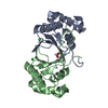
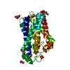
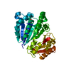
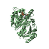
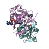
| Title | Crystal structure of a member of HIT family of proteins from bacillus subtilis | ||||||
 Components Components | Histidine triad protein | ||||||
 Keywords Keywords | CELL CYCLE / HIT protein / PKCI-1 / cell-cycle regulation / histidine triad / NYSGXRC / Structural Genomics / Protein Structure Initiative / T2097 / New York SGX Research Center for Structural Genomics / PSI | ||||||
| Function / homology |  Function and homology information Function and homology information | ||||||
| Biological species |  | ||||||
| Method |  X-RAY DIFFRACTION / SYNCHROTRON / SIRAS / Resolution: 2.3 Å X-RAY DIFFRACTION / SYNCHROTRON / SIRAS / Resolution: 2.3 Å | ||||||
 Authors Authors | Rajashankar, K.R. / Lima, C.D. / Burley, S.K. / New York SGX Research Center for Structural Genomics (NYSGXRC) | ||||||
 Citation Citation | Journal: To be Published Title: Crystal structure of a member of HIT family of proteins from bacillus subtilis Authors: Rajashankar, K.R. / Lima, C.D. | ||||||
| History |
|
- Structure visualization
| Structure viewer | Molecule: MolmilJmol/JSmol |
|---|
- Downloads & links
-Download
| PDBx/mmCIF format | 1y23.cif.gz | 153.6 KB | Display | PDBx/mmCIF format |
|---|---|---|---|---|
| PDB format | pdb1y23.ent.gz | 122.2 KB | Display | PDB format |
| PDBx/mmJSON format | 1y23.json.gz | Tree view | PDBx/mmJSON format | |
| Others |  Other downloads Other downloads |
-Validation report
| Arichive directory | https://data.pdbj.org/pub/pdb/validation_reports/y2/1y23ftp://data.pdbj.org/pub/pdb/validation_reports/y2/1y23 | HTTPS FTP |
|---|
-Related structure data
| Similar structure data | |
|---|---|
| Other databases |

-Links
 PDBj
PDBj


- Assembly
| Deposited unit | 
| ||||||||||
|---|---|---|---|---|---|---|---|---|---|---|---|
| 1 | 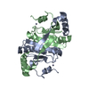
| ||||||||||
| 2 | 
| ||||||||||
| 3 | 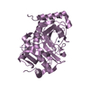
| ||||||||||
| Unit cell |
| ||||||||||
| Components on special symmetry positions |
| ||||||||||
| Details | Biological molecule is a dimer. Protomers A and B together form one biological assembly and so do protomers C and D. The partner protomer for molecule E is generated by the crystallographic two fold operation x, -y, -z+2/3 |
-Components
| #1: Protein | Mass: 16322.483 Da / Num. of mol.: 5 Source method: isolated from a genetically manipulated source Source: (gene. exp.) #2: Chemical | ChemComp-ZN /   Mass: 65.409 Da / Num. of mol.: 5 / Source method: obtained synthetically / Formula: Zn Mass: 65.409 Da / Num. of mol.: 5 / Source method: obtained synthetically / Formula: Zn#3: Chemical | ChemComp-MG / |   Mass: 24.305 Da / Num. of mol.: 1 / Source method: obtained synthetically / Formula: Mg Mass: 24.305 Da / Num. of mol.: 1 / Source method: obtained synthetically / Formula: Mg#4: Water | ChemComp-HOH / |  Mass: 18.015 Da / Num. of mol.: 287 / Source method: isolated from a natural source / Formula: H2O Mass: 18.015 Da / Num. of mol.: 287 / Source method: isolated from a natural source / Formula: H2O |
|---|
-Experimental details
-Experiment
| Experiment | Method: X-RAY DIFFRACTION / Number of used crystals: 1 |
|---|
- Sample preparation
| Crystal | Density Matthews: 2.38 Å3/Da / Density % sol: 47.98 % |
|---|---|
| Crystal grow | Temperature: 291 K / Method: vapor diffusion, hanging drop / pH: 5.5 Details: 19.5% Peg 4000, 0.5M Ammonium Acetate, 0.1M Sodium Citrate, pH 5.5, VAPOR DIFFUSION, HANGING DROP, temperature 291K |
-Data collection
| Diffraction | Mean temperature: 100 K |
|---|---|
| Diffraction source | Source: SYNCHROTRON / Site: NSLS  / Beamline: X4A / Wavelength: 0.98 Å / Beamline: X4A / Wavelength: 0.98 Å |
| Detector | Type: ADSC QUANTUM 4 / Detector: CCD |
| Radiation | Monochromator: SAGITALLY FOCUSED Si(111) / Protocol: SINGLE WAVELENGTH / Monochromatic (M) / Laue (L): M / Scattering type: x-ray |
| Radiation wavelength | Wavelength: 0.98 Å / Relative weight: 1 |
| Reflection | Resolution: 2.3→19.96 Å / Num. all: 35084 / Num. obs: 35084 / % possible obs: 99.6 % / Observed criterion σ(F): 0 / Observed criterion σ(I): 0 / Redundancy: 17.5 % / Biso Wilson estimate: 18.7 Å2 / Rsym value: 0.086 |
| Reflection shell | Resolution: 2.3→2.38 Å / Num. unique all: 3452 / Rsym value: 0.363 / % possible all: 99.8 |
- Processing
| Software |
| ||||||||||||||||||||||||||||||||||||
|---|---|---|---|---|---|---|---|---|---|---|---|---|---|---|---|---|---|---|---|---|---|---|---|---|---|---|---|---|---|---|---|---|---|---|---|---|---|
| Refinement | Method to determine structure: SIRAS Starting model: Experimental electron density map from Hg SIRAS density modified phases Resolution: 2.3→19.96 Å / Rfactor Rfree error: 0.006 / Data cutoff high absF: 248233.08 / Data cutoff low absF: 0 / Isotropic thermal model: RESTRAINED / Cross valid method: THROUGHOUT / σ(F): 0 / Stereochemistry target values: Engh & Huber
| ||||||||||||||||||||||||||||||||||||
| Solvent computation | Solvent model: FLAT MODEL / Bsol: 41.848 Å2 / ksol: 0.368286 e/Å3 | ||||||||||||||||||||||||||||||||||||
| Displacement parameters | Biso mean: 28.5 Å2
| ||||||||||||||||||||||||||||||||||||
| Refine analyze |
| ||||||||||||||||||||||||||||||||||||
| Refinement step | Cycle: LAST / Resolution: 2.3→19.96 Å
| ||||||||||||||||||||||||||||||||||||
| Refine LS restraints |
| ||||||||||||||||||||||||||||||||||||
| LS refinement shell | Resolution: 2.3→2.44 Å / Rfactor Rfree error: 0.018 / Total num. of bins used: 6
| ||||||||||||||||||||||||||||||||||||
| Xplor file |
|
