 Movie
Movie Controller

[English] 日本語
 Yorodumi
- PDB-1y0j: Zinc fingers as protein recognition motifs: structural basis for ... -
+ Open data

- Basic information
| Entry | Database: PDB / ID: 1y0j | ||||||
|---|---|---|---|---|---|---|---|
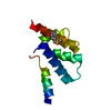
| Title | Zinc fingers as protein recognition motifs: structural basis for the GATA-1/Friend of GATA interaction | ||||||
 Components Components |
| ||||||
 Keywords Keywords | DNA BINDING PROTEIN / zinc finger / GATA-1 / FOG / protein-protein complex | ||||||
| Function / homology |  Function and homology information Function and homology informationlymph gland plasmatocyte differentiation / lymph gland crystal cell differentiation / negative regulation of hemocyte differentiation / amnioserosa maintenance / positive regulation of antibacterial peptide biosynthetic process / regulation of primitive erythrocyte differentiation / basophil differentiation / eosinophil fate commitment / regulation of definitive erythrocyte differentiation / germ-band shortening ...lymph gland plasmatocyte differentiation / lymph gland crystal cell differentiation / negative regulation of hemocyte differentiation / amnioserosa maintenance / positive regulation of antibacterial peptide biosynthetic process / regulation of primitive erythrocyte differentiation / basophil differentiation / eosinophil fate commitment / regulation of definitive erythrocyte differentiation / germ-band shortening / larval lymph gland hemopoiesis / lymph gland development / Factors involved in megakaryocyte development and platelet production / compound eye development / RUNX1 regulates genes involved in megakaryocyte differentiation and platelet function / regulation of glycoprotein biosynthetic process / megakaryocyte differentiation / chaeta development / RUNX1 regulates genes involved in megakaryocyte differentiation and platelet function / cell development / RUNX1 regulates transcription of genes involved in differentiation of HSCs / dendritic cell differentiation / Sertoli cell development / Factors involved in megakaryocyte development and platelet production / positive regulation of mast cell degranulation / cellular response to follicle-stimulating hormone stimulus / negative regulation of bone mineralization / myeloid cell differentiation / C2H2 zinc finger domain binding / embryonic hemopoiesis / platelet formation / DNA binding, bending / DNA-binding transcription repressor activity / positive regulation of osteoblast proliferation / animal organ regeneration / cell fate commitment / negative regulation of extrinsic apoptotic signaling pathway in absence of ligand / cis-regulatory region sequence-specific DNA binding / erythrocyte development / transcription repressor complex / negative regulation of insulin receptor signaling pathway / positive regulation of erythrocyte differentiation / homeostasis of number of cells within a tissue / cellular response to cAMP / transcription coregulator binding / erythrocyte differentiation / RNA polymerase II transcription regulatory region sequence-specific DNA binding / protein-DNA complex / chromatin DNA binding / platelet aggregation / male gonad development / transcription coactivator binding / sequence-specific double-stranded DNA binding / p53 binding / cell-cell signaling / heart development / cellular response to lipopolysaccharide / positive regulation of cytosolic calcium ion concentration / DNA-binding transcription activator activity, RNA polymerase II-specific / transcription regulator complex / sequence-specific DNA binding / in utero embryonic development / RNA polymerase II-specific DNA-binding transcription factor binding / DNA-binding transcription factor activity, RNA polymerase II-specific / cell differentiation / transcription cis-regulatory region binding / RNA polymerase II cis-regulatory region sequence-specific DNA binding / DNA-binding transcription factor activity / negative regulation of cell population proliferation / negative regulation of DNA-templated transcription / chromatin binding / regulation of DNA-templated transcription / negative regulation of apoptotic process / positive regulation of DNA-templated transcription / negative regulation of transcription by RNA polymerase II / positive regulation of transcription by RNA polymerase II / DNA binding / zinc ion binding / nucleoplasm / nucleus Similarity search - Function | ||||||
| Biological species |   | ||||||
| Method | SOLUTION NMR / simulated annealing, molecular dynamics torsion angle dynamics | ||||||
 Authors Authors | Liew, C.K. / Simpson, R.J.Y. / Kwan, A.H.Y. / Crofts, L.A. / Loughlin, F.E. / Matthews, J.M. / Crossley, M. / Mackay, J.P. | ||||||
 Citation Citation | Journal: Proc.Natl.Acad.Sci.Usa / Year: 2005 Title: Zinc fingers as protein recognition motifs: Structural basis for the GATA-1/Friend of GATA interaction Authors: Liew, C.K. / Simpson, R.J.Y. / Kwan, A.H.Y. / Crofts, L.A. / Loughlin, F.E. / Matthews, J.M. / Crossley, M. / Mackay, J.P. | ||||||
| History |
|
- Structure visualization
| Structure viewer | Molecule: MolmilJmol/JSmol |
|---|
- Downloads & links
-Download
| PDBx/mmCIF format | 1y0j.cif.gz | 449 KB | Display | PDBx/mmCIF format |
|---|---|---|---|---|
| PDB format | pdb1y0j.ent.gz | 370.1 KB | Display | PDB format |
| PDBx/mmJSON format | 1y0j.json.gz | Tree view | PDBx/mmJSON format | |
| Others |  Other downloads Other downloads |
-Validation report
| Arichive directory | https://data.pdbj.org/pub/pdb/validation_reports/y0/1y0jftp://data.pdbj.org/pub/pdb/validation_reports/y0/1y0j | HTTPS FTP |
|---|
-Related structure data
| Related structure data | |
|---|---|
| Similar structure data |


-Links
 PDBj
PDBj


- Assembly
| Deposited unit | 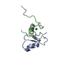
| |||||||||
|---|---|---|---|---|---|---|---|---|---|---|
| 1 |
| |||||||||
| NMR ensembles |
|
-Components
| #1: Protein/peptide | Mass: 5199.939 Da / Num. of mol.: 1 / Fragment: GNF Source method: isolated from a genetically manipulated source Source: (gene. exp.)  |
|---|---|
| #2: Protein/peptide | Mass: 3961.636 Da / Num. of mol.: 1 / Fragment: USF1 Source method: isolated from a genetically manipulated source Source: (gene. exp.) |
| #3: Chemical |   Mass: 65.409 Da / Num. of mol.: 2 / Source method: obtained synthetically / Formula: Zn Mass: 65.409 Da / Num. of mol.: 2 / Source method: obtained synthetically / Formula: Zn |
-Experimental details
-Experiment
| Experiment | Method: SOLUTION NMR | ||||||||||||||||||||||||||||||||||||||||||||
|---|---|---|---|---|---|---|---|---|---|---|---|---|---|---|---|---|---|---|---|---|---|---|---|---|---|---|---|---|---|---|---|---|---|---|---|---|---|---|---|---|---|---|---|---|---|
| NMR experiment |
| ||||||||||||||||||||||||||||||||||||||||||||
| NMR details | Text: Other commonly used standard triple-resonance NMR experiments were also recorded. |
 HNCA
HNCA- Sample preparation
| Details |
| |||||||||||||||
|---|---|---|---|---|---|---|---|---|---|---|---|---|---|---|---|---|
| Sample conditions | pH: 5.5 / Pressure: ambient / Temperature: 280 K |
-NMR measurement
| Radiation | Protocol: SINGLE WAVELENGTH / Monochromatic (M) / Laue (L): M |
|---|---|
| Radiation wavelength | Relative weight: 1 |
| NMR spectrometer | Type: Bruker DRX / Manufacturer: Bruker / Model: DRX / Field strength: 600 MHz |
- Processing
| NMR software |
| ||||||||||||||||||||||||
|---|---|---|---|---|---|---|---|---|---|---|---|---|---|---|---|---|---|---|---|---|---|---|---|---|---|
| Refinement | Method: simulated annealing, molecular dynamics torsion angle dynamics Software ordinal: 1 Details: Standard triple-resonance NMR spectroscopy was first used to determine the preliminary structure of the complex. Since the backbone folds of both GNF and USF1 did not appear to be ...Details: Standard triple-resonance NMR spectroscopy was first used to determine the preliminary structure of the complex. Since the backbone folds of both GNF and USF1 did not appear to be substantially altered to those of the proteins in isolation, the computer program Haddock was used to dock the two partner proteins using intermolecular NOEs obtained from double half-filtered NOESY experiments, and ambiguous restraints derived from mutagenesis and NMR titration experiments. | ||||||||||||||||||||||||
| NMR representative | Selection criteria: lowest energy | ||||||||||||||||||||||||
| NMR ensemble | Conformer selection criteria: structures with the lowest energy Conformers calculated total number: 100 / Conformers submitted total number: 20 |
