 Movie
Movie Controller

[English] 日本語
 Yorodumi
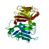
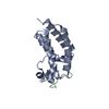
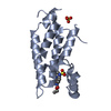
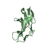
- PDB-1xt5: Crystal Structure of VCBP3, domain 1, from Branchiostoma floridae -
+ Open data

- Basic information
| Entry | Database: PDB / ID: 1xt5 | ||||||
|---|---|---|---|---|---|---|---|
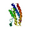
| Title | Crystal Structure of VCBP3, domain 1, from Branchiostoma floridae | ||||||
 Components Components | variable region-containing chitin-binding protein 3 | ||||||
 Keywords Keywords | IMMUNE SYSTEM / innate immunity / vcbp / primordial antigen receptor / florida lancelet / amphioxus | ||||||
| Function / homology |  Function and homology information Function and homology informationendochitinase activity / chitinase / chitin catabolic process / chitin binding / extracellular region Similarity search - Function | ||||||
| Biological species |  | ||||||
| Method |  X-RAY DIFFRACTION / SYNCHROTRON / MAD / Resolution: 1.15 Å X-RAY DIFFRACTION / SYNCHROTRON / MAD / Resolution: 1.15 Å | ||||||
 Authors Authors | Hernandez Prada, J.A. / Haire, R.N. / Cannon, J.P. / Allaire, M. / Jakoncic, J. / Stojanoff, V. / Litman, G.W. / Ostrov, D.A. | ||||||
 Citation Citation | Journal: Nat.Immunol. / Year: 2006 Title: Ancient evolutionary origin of diversified variable regions demonstrated by crystal structures of an immune-type receptor in amphioxus. Authors: Haire, R.N. / Allaire, M. / Jakoncic, J. / Stojanoff, V. / Cannon, J.P. / Litman, G.W. / Ostrov, D.A. #1: Journal: Acta Crystallogr.,Sect.D / Year: 2004Title: Crystallization and preliminary X-ray analysis of VCBP3 from Branchiostoma floridae. Authors: Hernandez Prada, J.A. / Haire, R.N. / Cannon, J.P. / Litman, G.W. / Ostrov, D.A. | ||||||
| History |
|
- Structure visualization
| Structure viewer | Molecule: MolmilJmol/JSmol |
|---|
- Downloads & links
-Download
| PDBx/mmCIF format | 1xt5.cif.gz | 76.2 KB | Display | PDBx/mmCIF format |
|---|---|---|---|---|
| PDB format | pdb1xt5.ent.gz | 56.4 KB | Display | PDB format |
| PDBx/mmJSON format | 1xt5.json.gz | Tree view | PDBx/mmJSON format | |
| Others |  Other downloads Other downloads |
-Validation report
| Arichive directory | https://data.pdbj.org/pub/pdb/validation_reports/xt/1xt5ftp://data.pdbj.org/pub/pdb/validation_reports/xt/1xt5 | HTTPS FTP |
|---|
-Related structure data


-Links
 PDBj
PDBj- Assembly
| Deposited unit | 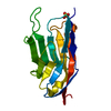
| |||||||||
|---|---|---|---|---|---|---|---|---|---|---|
| 1 |
| |||||||||
| Unit cell |
| |||||||||
| Components on special symmetry positions |
| |||||||||
| Details | biological unit unknown |
-Components
| #1: Protein | Mass: 14797.479 Da / Num. of mol.: 1 Fragment: sequence database residues 16-150: contains immunoglobulin like region (residues 33-146) Source method: isolated from a genetically manipulated source Source: (gene. exp.) Gene: VCBP3 / Production host:  |
|---|---|
| #2: Chemical | ChemComp-SO4 /   Mass: 96.063 Da / Num. of mol.: 1 / Source method: obtained synthetically / Formula: SO4 Mass: 96.063 Da / Num. of mol.: 1 / Source method: obtained synthetically / Formula: SO4 |
| #3: Water | ChemComp-HOH /  Mass: 18.015 Da / Num. of mol.: 223 / Source method: isolated from a natural source / Formula: H2O Mass: 18.015 Da / Num. of mol.: 223 / Source method: isolated from a natural source / Formula: H2O |
| Has protein modification | Y |
-Experimental details
-Experiment
| Experiment | Method: X-RAY DIFFRACTION / Number of used crystals: 1 |
|---|
- Sample preparation
| Crystal | Density Matthews: 2.708 Å3/Da / Density % sol: 54.57 % |
|---|---|
| Crystal grow | Temperature: 295 K / Method: vapor diffusion, hanging drop / pH: 8.5 Details: 1.2-1.4 M ammonium sulfate, 0.1 M NaCl, 0.1 M tris-HCl (or HEPES), 12% glycerol, pH 8.5, VAPOR DIFFUSION, HANGING DROP, temperature 295K |
-Data collection
| Diffraction | Mean temperature: 100 K | |||||||||||||||
|---|---|---|---|---|---|---|---|---|---|---|---|---|---|---|---|---|
| Diffraction source | Source: SYNCHROTRON / Site: NSLS  / Beamline: X6A / Wavelength: 0.8 / Wavelength: 0.9796, 0.97908, 0.95007 / Beamline: X6A / Wavelength: 0.8 / Wavelength: 0.9796, 0.97908, 0.95007 | |||||||||||||||
| Detector | Type: ADSC QUANTUM 210 / Detector: CCD / Date: Jun 13, 2004 Details: Oxford Danfysik toroidal focusing mirror, Si(111) channel cut monochromator | |||||||||||||||
| Radiation | Monochromator: Si(111) channel cut monochromator / Protocol: MAD / Monochromatic (M) / Laue (L): M / Scattering type: x-ray | |||||||||||||||
| Radiation wavelength |
| |||||||||||||||
| Reflection | Resolution: 1.15→30 Å / Num. all: 57420 / Num. obs: 57420 / % possible obs: 99.8 % / Observed criterion σ(F): 0 / Observed criterion σ(I): 0 / Redundancy: 14 % / Rmerge(I) obs: 0.049 / Net I/σ(I): 59.3 | |||||||||||||||
| Reflection shell | Resolution: 1.15→1.16 Å / Rmerge(I) obs: 0.37 / Mean I/σ(I) obs: 6 / Num. unique all: 1898 / % possible all: 99.7 |
- Processing
| Software |
| |||||||||||||||||||||||||||||||||||
|---|---|---|---|---|---|---|---|---|---|---|---|---|---|---|---|---|---|---|---|---|---|---|---|---|---|---|---|---|---|---|---|---|---|---|---|---|
| Refinement | Method to determine structure: MAD / Resolution: 1.15→20 Å / Num. parameters: 11572 / Num. restraintsaints: 13947 / Isotropic thermal model: Anisotropic / Cross valid method: FREE R / σ(F): 0 / σ(I): 0 / Stereochemistry target values: ENGH AND HUBER Details: ANISOTROPIC SCALING APPLIED BY THE METHOD OF PARKIN, MOEZZI & HOPE, J.APPL.CRYST.28(1995)53-56 ANISOTROPIC REFINEMENT REDUCED FREE R (NO CUTOFF) BY ~3%.
| |||||||||||||||||||||||||||||||||||
| Refine analyze | Num. disordered residues: 5 / Occupancy sum hydrogen: 904.7 / Occupancy sum non hydrogen: 1214.85 | |||||||||||||||||||||||||||||||||||
| Refinement step | Cycle: LAST / Resolution: 1.15→20 Å
| |||||||||||||||||||||||||||||||||||
| Refine LS restraints |
| |||||||||||||||||||||||||||||||||||
| LS refinement shell |
|
