 Movie
Movie Controller

[English] 日本語
 Yorodumi
- PDB-1xsv: X-ray crystal structure of conserved hypothetical UPF0122 protein... -
+ Open data

- Basic information
| Entry | Database: PDB / ID: 1xsv | ||||||
|---|---|---|---|---|---|---|---|
| Title | X-ray crystal structure of conserved hypothetical UPF0122 protein SAV1236 from Staphylococcus aureus subsp. aureus Mu50 | ||||||
 Components Components | Hypothetical UPF0122 protein SAV1236 | ||||||
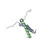
 Keywords Keywords | UNKNOWN FUNCTION / helix-turn-helix / putative DNA-binding protein / signal recognition particle / UPF0122 | ||||||
| Function / homology |  Function and homology information Function and homology informationPutative helix-turn-helix protein, YlxM/p13-like / : / Putative helix-turn-helix protein, YlxM / p13 like / RNA polymerase sigma factor, region 3/4-like / Winged helix-like DNA-binding domain superfamily/Winged helix DNA-binding domain / Arc Repressor Mutant, subunit A / Winged helix-like DNA-binding domain superfamily / Orthogonal Bundle / Mainly Alpha Similarity search - Domain/homology | ||||||
| Biological species |   Staphylococcus aureus subsp. aureus (bacteria) Staphylococcus aureus subsp. aureus (bacteria) | ||||||
| Method |  X-RAY DIFFRACTION / SYNCHROTRON / SAD / Resolution: 1.7 Å X-RAY DIFFRACTION / SYNCHROTRON / SAD / Resolution: 1.7 Å | ||||||
 Authors Authors | Walker, J.R. / Xu, X. / Virag, C. / McDonald, M.-L. / Houston, S. / Buzadzija, K. / Vedadi, M. / Dharamsi, A. / Fiebig, K.M. / Savchenko, A. | ||||||
 Citation Citation | Journal: To be Published Title: 1.7 Angstrom Crystal Structure of Conserved Hypothetical UPF0122 Protein SAV1236 From Staphylococcus aureus Authors: Walker, J.R. / Xu, X. / Virag, C. / McDonald, M.-L. / Houston, S. / Buzadzija, K. / Vedadi, M. / Dharamsi, A. / Fiebig, K.M. / Savchenko, A. | ||||||
| History |
|
- Structure visualization
| Structure viewer | Molecule: MolmilJmol/JSmol |
|---|
- Downloads & links
-Download
| PDBx/mmCIF format | 1xsv.cif.gz | 63.5 KB | Display | PDBx/mmCIF format |
|---|---|---|---|---|
| PDB format | pdb1xsv.ent.gz | 47.1 KB | Display | PDB format |
| PDBx/mmJSON format | 1xsv.json.gz | Tree view | PDBx/mmJSON format | |
| Others |  Other downloads Other downloads |
-Validation report
| Arichive directory | https://data.pdbj.org/pub/pdb/validation_reports/xs/1xsvftp://data.pdbj.org/pub/pdb/validation_reports/xs/1xsv | HTTPS FTP |
|---|
-Related structure data
| Similar structure data |
|---|


-Links
 PDBj
PDBj
- Assembly
| Deposited unit | 
| ||||||||
|---|---|---|---|---|---|---|---|---|---|
| 1 |
| ||||||||
| Unit cell |
| ||||||||
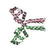
| Details | The biological assembly is a dimer, consisting of Chains A and B. |
-Components
| #1: Protein | Mass: 13972.197 Da / Num. of mol.: 2 Source method: isolated from a genetically manipulated source Source: (gene. exp.) Staphylococcus aureus subsp. aureus (bacteria)Species: Staphylococcus aureus / Strain: subsp. aureus / Gene: SAV1236 / Plasmid: pPW2 / Production host: #2: Water | ChemComp-HOH / |  Mass: 18.015 Da / Num. of mol.: 272 / Source method: isolated from a natural source / Formula: H2O Mass: 18.015 Da / Num. of mol.: 272 / Source method: isolated from a natural source / Formula: H2OHas protein modification | Y | |
|---|
-Experimental details
-Experiment
| Experiment | Method: X-RAY DIFFRACTION / Number of used crystals: 1 |
|---|
- Sample preparation
| Crystal | Density Matthews: 2.31 Å3/Da / Density % sol: 46.8 % |
|---|---|
| Crystal grow | Temperature: 298 K / Method: vapor diffusion, hanging drop / pH: 4.6 Details: 0.1M Sodium Acetate, 2.0M Ammonium sulfate, pH 4.6, VAPOR DIFFUSION, HANGING DROP, temperature 298K |
-Data collection
| Diffraction | Mean temperature: 100 K |
|---|---|
| Diffraction source | Source: SYNCHROTRON / Site: APS  / Beamline: 17-ID / Wavelength: 0.97936 Å / Beamline: 17-ID / Wavelength: 0.97936 Å |
| Detector | Type: ADSC QUANTUM 210 / Detector: CCD / Date: Aug 6, 2004 |
| Radiation | Monochromator: Si(111) double crystal / Protocol: SINGLE WAVELENGTH / Monochromatic (M) / Laue (L): M / Scattering type: x-ray |
| Radiation wavelength | Wavelength: 0.97936 Å / Relative weight: 1 |
| Reflection | Resolution: 1.7→32 Å / Num. all: 24963 / Num. obs: 24963 / % possible obs: 99.4 % / Observed criterion σ(F): 0 / Observed criterion σ(I): -3 / Redundancy: 5.68 % / Biso Wilson estimate: 19 Å2 / Rmerge(I) obs: 0.054 |
| Reflection shell | Resolution: 1.7→1.78 Å / Redundancy: 5.9 % / Rmerge(I) obs: 0.23 / % possible all: 100 |
- Processing
| Software |
| ||||||||||||||||||||||||||||||||||||
|---|---|---|---|---|---|---|---|---|---|---|---|---|---|---|---|---|---|---|---|---|---|---|---|---|---|---|---|---|---|---|---|---|---|---|---|---|---|
| Refinement | Method to determine structure: SAD / Resolution: 1.7→32.54 Å / Rfactor Rfree error: 0.006 / Data cutoff high absF: 1237412.1 / Data cutoff low absF: 0 / Isotropic thermal model: RESTRAINED / Cross valid method: THROUGHOUT / σ(F): 0 / Stereochemistry target values: Engh & Huber
| ||||||||||||||||||||||||||||||||||||
| Solvent computation | Solvent model: FLAT MODEL / Bsol: 52.5857 Å2 / ksol: 0.409932 e/Å3 | ||||||||||||||||||||||||||||||||||||
| Displacement parameters | Biso mean: 21.1 Å2
| ||||||||||||||||||||||||||||||||||||
| Refine analyze |
| ||||||||||||||||||||||||||||||||||||
| Refinement step | Cycle: LAST / Resolution: 1.7→32.54 Å
| ||||||||||||||||||||||||||||||||||||
| Refine LS restraints |
| ||||||||||||||||||||||||||||||||||||
| LS refinement shell | Resolution: 1.7→1.81 Å / Rfactor Rfree error: 0.017 / Total num. of bins used: 6
| ||||||||||||||||||||||||||||||||||||
| Xplor file |
|
