 Movie
Movie Controller

[English] 日本語
 Yorodumi
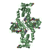
- PDB-1s7o: Crystal structure of putative DNA binding protein SP_1288 from St... -
+ Open data

- Basic information
| Entry | Database: PDB / ID: 1s7o | ||||||
|---|---|---|---|---|---|---|---|
| Title | Crystal structure of putative DNA binding protein SP_1288 from Streptococcus pygenes | ||||||
 Components Components | Hypothetical UPF0122 protein SPy1201/SpyM3_0842/SPs1042/spyM18_1152 | ||||||
 Keywords Keywords | STRUCTURAL GENOMICS / UNKNOWN FUNCTION / Putative DNA binding protein / Signal recognition particle / RNA polymerase Sigma Factor / X-Rat crystallography / UPF0122 / structure funded by NIH / PSI / Protein Structure Initiative / Berkeley Structural Genomics Center / BSGC | ||||||
| Function / homology |  Function and homology information Function and homology informationPutative helix-turn-helix protein, YlxM/p13-like / : / Putative helix-turn-helix protein, YlxM / p13 like / RNA polymerase sigma factor, region 3/4-like / Winged helix-like DNA-binding domain superfamily/Winged helix DNA-binding domain / Arc Repressor Mutant, subunit A / Winged helix-like DNA-binding domain superfamily / Orthogonal Bundle / Mainly Alpha Similarity search - Domain/homology | ||||||
| Biological species |  Streptococcus pyogenes serotype M3 (bacteria) Streptococcus pyogenes serotype M3 (bacteria) | ||||||
| Method |  X-RAY DIFFRACTION / SYNCHROTRON / SAD / Resolution: 2.31 Å X-RAY DIFFRACTION / SYNCHROTRON / SAD / Resolution: 2.31 Å | ||||||
 Authors Authors | Oganesyan, V. / Pufan, R. / DeGiovanni, A. / Yokota, H. / Kim, R. / Kim, S.-H. / Berkeley Structural Genomics Center (BSGC) | ||||||
 Citation Citation | Journal: Acta Crystallogr.,Sect.D / Year: 2004 Title: Structure of the putative DNA-binding protein SP_1288 from Streptococcus pyogenes. Authors: Oganesyan, V. / Pufan, R. / DeGiovanni, A. / Yokota, H. / Kim, R. / Kim, S.H. | ||||||
| History |
|
- Structure visualization
| Structure viewer | Molecule: MolmilJmol/JSmol |
|---|
- Downloads & links
-Download
| PDBx/mmCIF format | 1s7o.cif.gz | 77.8 KB | Display | PDBx/mmCIF format |
|---|---|---|---|---|
| PDB format | pdb1s7o.ent.gz | 61.2 KB | Display | PDB format |
| PDBx/mmJSON format | 1s7o.json.gz | Tree view | PDBx/mmJSON format | |
| Others |  Other downloads Other downloads |
-Validation report
| Arichive directory | https://data.pdbj.org/pub/pdb/validation_reports/s7/1s7oftp://data.pdbj.org/pub/pdb/validation_reports/s7/1s7o | HTTPS FTP |
|---|
-Related structure data
| Similar structure data | |
|---|---|
| Other databases |


-Links
 PDBj
PDBj
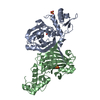
- Assembly
| Deposited unit | 
| ||||||||
|---|---|---|---|---|---|---|---|---|---|
| 1 |
| ||||||||
| 2 | 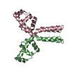
| ||||||||
| 3 | 
| ||||||||
| Unit cell |
| ||||||||
| Details | The biological assembly is probably dimer; molecules with chain identifiers B and C |
-Components
| #1: Protein | Mass: 13604.293 Da / Num. of mol.: 3 / Fragment: gi 15675166 Source method: isolated from a genetically manipulated source Source: (gene. exp.) Streptococcus pyogenes serotype M3 (bacteria)Species: Streptococcus pyogenes / Strain: serotype M3 / Gene: SPY1201, SPYM3_0842, SPS1042, SPYM18_1152 / Plasmid: pB3.1325B / Production host: #2: Water | ChemComp-HOH / |  Mass: 18.015 Da / Num. of mol.: 103 / Source method: isolated from a natural source / Formula: H2O Mass: 18.015 Da / Num. of mol.: 103 / Source method: isolated from a natural source / Formula: H2O |
|---|
-Experimental details
-Experiment
| Experiment | Method: X-RAY DIFFRACTION / Number of used crystals: 1 |
|---|
- Sample preparation
| Crystal | Density Matthews: 3.19 Å3/Da / Density % sol: 61.17 % |
|---|---|
| Crystal grow | Temperature: 293 K / Method: vapor diffusion, hanging drop / pH: 3.2 Details: Citric Acid, Ammonium Nitrate, pH 3.2, VAPOR DIFFUSION, HANGING DROP, temperature 293.0K |
-Data collection
| Diffraction | Mean temperature: 100 K |
|---|---|
| Diffraction source | Source: SYNCHROTRON / Site: ALS  / Beamline: 5.0.2 / Wavelength: 0.9794 Å / Beamline: 5.0.2 / Wavelength: 0.9794 Å |
| Detector | Type: ADSC QUANTUM 4 / Detector: CCD / Date: Nov 8, 2003 / Details: Mirrors |
| Radiation | Monochromator: Double-crystal, Si (111) / Protocol: SINGLE WAVELENGTH / Monochromatic (M) / Laue (L): M / Scattering type: x-ray |
| Radiation wavelength | Wavelength: 0.9794 Å / Relative weight: 1 |
| Reflection | Resolution: 2.3→38 Å / Num. all: 40688 / Num. obs: 40607 / % possible obs: 99.8 % / Observed criterion σ(F): 0 / Observed criterion σ(I): 0 / Redundancy: 4 % / Biso Wilson estimate: 39 Å2 / Rmerge(I) obs: 0.066 / Rsym value: 0.066 / Net I/σ(I): 19 |
| Reflection shell | Resolution: 2.3→2.38 Å / Redundancy: 4 % / Rmerge(I) obs: 0.336 / Mean I/σ(I) obs: 4 / Num. unique all: 4066 / Rsym value: 0.336 / % possible all: 99.8 |
- Processing
| Software |
| |||||||||||||||||||||||||||||||||||||||||||||||||||||||||||||||||||||||||||||||||||||||||||||||||||||||||||||||||||||||||||||||||||||||||||||||||||||||||||||||||||||||||||||||
|---|---|---|---|---|---|---|---|---|---|---|---|---|---|---|---|---|---|---|---|---|---|---|---|---|---|---|---|---|---|---|---|---|---|---|---|---|---|---|---|---|---|---|---|---|---|---|---|---|---|---|---|---|---|---|---|---|---|---|---|---|---|---|---|---|---|---|---|---|---|---|---|---|---|---|---|---|---|---|---|---|---|---|---|---|---|---|---|---|---|---|---|---|---|---|---|---|---|---|---|---|---|---|---|---|---|---|---|---|---|---|---|---|---|---|---|---|---|---|---|---|---|---|---|---|---|---|---|---|---|---|---|---|---|---|---|---|---|---|---|---|---|---|---|---|---|---|---|---|---|---|---|---|---|---|---|---|---|---|---|---|---|---|---|---|---|---|---|---|---|---|---|---|---|---|---|---|
| Refinement | Method to determine structure: SAD / Resolution: 2.31→38.35 Å / Cor.coef. Fo:Fc: 0.933 / Cor.coef. Fo:Fc free: 0.911 / SU B: 6.301 / SU ML: 0.157 / TLS residual ADP flag: LIKELY RESIDUAL / Cross valid method: THROUGHOUT / σ(F): 0 / σ(I): 0 / ESU R: 0.281 / ESU R Free: 0.211 / Stereochemistry target values: MAXIMUM LIKELIHOOD
| |||||||||||||||||||||||||||||||||||||||||||||||||||||||||||||||||||||||||||||||||||||||||||||||||||||||||||||||||||||||||||||||||||||||||||||||||||||||||||||||||||||||||||||||
| Solvent computation | Ion probe radii: 0.8 Å / Shrinkage radii: 0.8 Å / VDW probe radii: 1.4 Å / Solvent model: BABINET MODEL WITH MASK | |||||||||||||||||||||||||||||||||||||||||||||||||||||||||||||||||||||||||||||||||||||||||||||||||||||||||||||||||||||||||||||||||||||||||||||||||||||||||||||||||||||||||||||||
| Displacement parameters | Biso mean: 27.264 Å2
| |||||||||||||||||||||||||||||||||||||||||||||||||||||||||||||||||||||||||||||||||||||||||||||||||||||||||||||||||||||||||||||||||||||||||||||||||||||||||||||||||||||||||||||||
| Refinement step | Cycle: LAST / Resolution: 2.31→38.35 Å
| |||||||||||||||||||||||||||||||||||||||||||||||||||||||||||||||||||||||||||||||||||||||||||||||||||||||||||||||||||||||||||||||||||||||||||||||||||||||||||||||||||||||||||||||
| Refine LS restraints |
| |||||||||||||||||||||||||||||||||||||||||||||||||||||||||||||||||||||||||||||||||||||||||||||||||||||||||||||||||||||||||||||||||||||||||||||||||||||||||||||||||||||||||||||||
| LS refinement shell | Resolution: 2.308→2.368 Å / Rfactor Rfree error: 0.02 / Total num. of bins used: 20
| |||||||||||||||||||||||||||||||||||||||||||||||||||||||||||||||||||||||||||||||||||||||||||||||||||||||||||||||||||||||||||||||||||||||||||||||||||||||||||||||||||||||||||||||
| Refinement TLS params. | Method: refined / Refine-ID: X-RAY DIFFRACTION
| |||||||||||||||||||||||||||||||||||||||||||||||||||||||||||||||||||||||||||||||||||||||||||||||||||||||||||||||||||||||||||||||||||||||||||||||||||||||||||||||||||||||||||||||
| Refinement TLS group |
|
