 Movie
Movie Controller

[English] 日本語
 Yorodumi
- PDB-1xso: THREE-DIMENSIONAL STRUCTURE OF XENOPUS LAEVIS CU,ZN SUPEROXIDE DI... -
+ Open data

- Basic information
| Entry | Database: PDB / ID: 1xso | ||||||
|---|---|---|---|---|---|---|---|
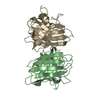
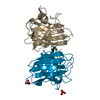
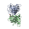
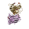
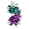
| Title | THREE-DIMENSIONAL STRUCTURE OF XENOPUS LAEVIS CU,ZN SUPEROXIDE DISMUTASE B DETERMINED BY X-RAY CRYSTALLOGRAPHY AT 1.5 ANGSTROMS RESOLUTION | ||||||
 Components Components | COPPER,ZINC SUPEROXIDE DISMUTASE | ||||||
 Keywords Keywords | OXIDOREDUCTASE (SUPEROXIDE ACCEPTOR) | ||||||
| Function / homology |  Function and homology information Function and homology informationOxidoreductases; Acting on a sulfur group of donors / superoxide dismutase / superoxide dismutase activity / removal of superoxide radicals / peroxisome / copper ion binding / mitochondrion / nucleus / cytosol Similarity search - Function | ||||||
| Biological species | |||||||
| Method |  X-RAY DIFFRACTION / SYNCHROTRON / Resolution: 1.49 Å X-RAY DIFFRACTION / SYNCHROTRON / Resolution: 1.49 Å | ||||||
 Authors Authors | Djinovic Carugo, K. / Coda, A. / Battistoni, A. / Carri, M.T. / Polticelli, F. / Desideri, A. / Rotilio, G. / Wilson, K.S. / Bolognesi, M. | ||||||
 Citation Citation | Journal: Acta Crystallogr.,Sect.D / Year: 1996 Title: Three-dimensional structure of Xenopus laevis Cu,Zn superoxide dismutase b determined by X-ray crystallography at 1.5 A resolution. Authors: Djinovic Carugo, K. / Battistoni, A. / Carri, M.T. / Polticelli, F. / Desideri, A. / Rotilio, G. / Coda, A. / Wilson, K.S. / Bolognesi, M. #1: Journal: J.Mol.Biol. / Year: 1994Title: Conserved Patterns in the Cu,Zn Superoxide Dismutase Family Authors: Bordo, D. / Djinovic, K. / Bolognesi, K.M. #2: Journal: J.Mol.Biol. / Year: 1994Title: Crystallographic Study of Azide-Inhibited Bovine Cu,Zn Superoxide Dismutase Authors: Djinovic Carugo, K. / Polticelli, K.F. / Desideri, A. / Rotilio, G. / Wilson, K.S. / Bolognesi, M. #3: Journal: FEBS Lett. / Year: 1994Title: Crystal Structure of the Cyanide-Inhibited Xenopus Laevis Cu,Zn Superoxide Dismutase at 98 K Authors: Djinovic Carugo, K. / Battistoni, K.A. / Carri, M.T. / Polticelli, F. / Desideri, A. / Rotilio, G. / Coda, A. / Bolognesi, M. #4: Journal: Biochem.Biophys.Res.Commun. / Year: 1993Title: Crystallisation and Preliminary Crystallographic Analysis of Recombinant Xenopus Laevis Cu,Zn Superoxide Dismutase B Authors: Djinovic Carugo, K. / Collyer, K.C. / Coda, A. / Carri, M.T. / Battistoni, A. / Botaro, G. / Polticelli, F. / Desideri, A. / Bolognesi, M. #5: Journal: J.Mol.Biol. / Year: 1992Title: Crystal Structure of Yeast Cu,Zn Superoxide Dismutase Authors: Djinovic, K. / Gatti, G. / Antolini, L. / Pelosi, G. / Desideri, A. / Falconi, M. / Marmocchi, F. / Rotilio, G. / Bolognesi, M. | ||||||
| History |
|
- Structure visualization
| Structure viewer | Molecule: MolmilJmol/JSmol |
|---|
- Downloads & links
-Download
| PDBx/mmCIF format | 1xso.cif.gz | 77.2 KB | Display | PDBx/mmCIF format |
|---|---|---|---|---|
| PDB format | pdb1xso.ent.gz | 56.7 KB | Display | PDB format |
| PDBx/mmJSON format | 1xso.json.gz | Tree view | PDBx/mmJSON format | |
| Others |  Other downloads Other downloads |
-Validation report
| Arichive directory | https://data.pdbj.org/pub/pdb/validation_reports/xs/1xsoftp://data.pdbj.org/pub/pdb/validation_reports/xs/1xso | HTTPS FTP |
|---|
-Related structure data
| Similar structure data |
|---|


-Links
 PDBj
PDBj
- Assembly
| Deposited unit | 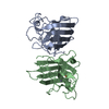
| ||||||||
|---|---|---|---|---|---|---|---|---|---|
| 1 |
| ||||||||
| Unit cell |
| ||||||||
| Noncrystallographic symmetry (NCS) | NCS oper: (Code: given Matrix: (0.98783, -0.01029, -0.15522), Vector: Details | MTRIX THE TRANSFORMATIONS PRESENTED ON MTRIX RECORDS BELOW DESCRIBE NON-CRYSTALLOGRAPHIC RELATIONSHIPS AMONG THE VARIOUS DOMAINS IN THIS ENTRY. APPLYING THE APPROPRIATE MTRIX TRANSFORMATION TO THE RESIDUES LISTED FIRST WILL YIELD APPROXIMATE COORDINATES FOR THE RESIDUES LISTED SECOND. APPLIED TO TRANSFORMED TO MTRIX RESIDUES RESIDUES RMSD M1 A 2 .. A 151 B 2 .. B 151 0.279 CALCULATED FOR C-ALPHA ATOMS | |
-Components
| #1: Protein | Mass: 15306.876 Da / Num. of mol.: 2 Source method: isolated from a genetically manipulated source Source: (gene. exp.)  #2: Chemical |   Mass: 63.546 Da / Num. of mol.: 2 / Source method: obtained synthetically / Formula: Cu Mass: 63.546 Da / Num. of mol.: 2 / Source method: obtained synthetically / Formula: Cu#3: Chemical |   Mass: 65.409 Da / Num. of mol.: 2 / Source method: obtained synthetically / Formula: Zn Mass: 65.409 Da / Num. of mol.: 2 / Source method: obtained synthetically / Formula: Zn#4: Water | ChemComp-HOH / |  Mass: 18.015 Da / Num. of mol.: 350 / Source method: isolated from a natural source / Formula: H2O Mass: 18.015 Da / Num. of mol.: 350 / Source method: isolated from a natural source / Formula: H2OHas protein modification | Y | |
|---|
-Experimental details
-Experiment
| Experiment | Method: X-RAY DIFFRACTION |
|---|
- Sample preparation
| Crystal | Density Matthews: 2.43 Å3/Da / Density % sol: 49.36 % | ||||||||||||||||||||
|---|---|---|---|---|---|---|---|---|---|---|---|---|---|---|---|---|---|---|---|---|---|
| Crystal grow | *PLUS Temperature: 301 K / pH: 6 / Method: vapor diffusion, sitting drop | ||||||||||||||||||||
| Components of the solutions | *PLUS
|
-Data collection
| Diffraction source | Source: SYNCHROTRON / Site: EMBL/DESY, HAMBURG  / Beamline: X31 / Wavelength: 0.92 / Beamline: X31 / Wavelength: 0.92 |
|---|---|
| Detector | Type: CUSTOM-MADE / Detector: IMAGE PLATE / Date: Aug 18, 1993 |
| Radiation | Scattering type: x-ray |
| Radiation wavelength | Wavelength: 0.92 Å / Relative weight: 1 |
| Reflection | Num. obs: 49209 / % possible obs: 98.8 % / Observed criterion σ(I): 1 / Redundancy: 4.98 % / Rmerge(I) obs: 0.078 |
| Reflection | *PLUS Highest resolution: 1.49 Å / Num. measured all: 245181 / Rmerge(I) obs: 0.078 |
- Processing
| Software |
| ||||||||||||||||||||||||||||||||||||||||||||||||||||||||||||||||||||||||||||||||
|---|---|---|---|---|---|---|---|---|---|---|---|---|---|---|---|---|---|---|---|---|---|---|---|---|---|---|---|---|---|---|---|---|---|---|---|---|---|---|---|---|---|---|---|---|---|---|---|---|---|---|---|---|---|---|---|---|---|---|---|---|---|---|---|---|---|---|---|---|---|---|---|---|---|---|---|---|---|---|---|---|---|
| Refinement | Resolution: 1.49→10 Å / σ(F): 0 / Details: RESIDUE 24 IS IN POOR ELECTRON DENSITY.
| ||||||||||||||||||||||||||||||||||||||||||||||||||||||||||||||||||||||||||||||||
| Displacement parameters | Biso mean: 20.3 Å2 | ||||||||||||||||||||||||||||||||||||||||||||||||||||||||||||||||||||||||||||||||
| Refinement step | Cycle: LAST / Resolution: 1.49→10 Å
| ||||||||||||||||||||||||||||||||||||||||||||||||||||||||||||||||||||||||||||||||
| Refine LS restraints |
| ||||||||||||||||||||||||||||||||||||||||||||||||||||||||||||||||||||||||||||||||
| Refine LS restraints | *PLUS
|
