 Movie
Movie Controller


+ Open data

- Basic information
| Entry | Database: PDB / ID: 1xrx | ||||||
|---|---|---|---|---|---|---|---|
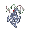
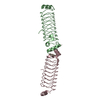
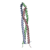
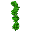
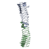
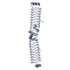
| Title | Crystal structure of a DNA-binding protein | ||||||
 Components Components | SeqA protein | ||||||
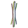
 Keywords Keywords | REPLICATION INHIBITOR / Protein filament / Left-handed helix / DNA replication inhibitor | ||||||
| Function / homology |  Function and homology information Function and homology informationSeqA-DNA complex / double-stranded methylated DNA binding / hemi-methylated DNA-binding / sister chromatid cohesion / DNA replication origin binding / negative regulation of DNA-templated DNA replication initiation / response to radiation / regulation of DNA-templated transcription / protein homodimerization activity / protein-containing complex ...SeqA-DNA complex / double-stranded methylated DNA binding / hemi-methylated DNA-binding / sister chromatid cohesion / DNA replication origin binding / negative regulation of DNA-templated DNA replication initiation / response to radiation / regulation of DNA-templated transcription / protein homodimerization activity / protein-containing complex / identical protein binding / cytosol Similarity search - Function | ||||||
| Biological species |  | ||||||
| Method |  X-RAY DIFFRACTION / SYNCHROTRON / MAD / Resolution: 2.15 Å X-RAY DIFFRACTION / SYNCHROTRON / MAD / Resolution: 2.15 Å | ||||||
 Authors Authors | Guarne, A. / Brendler, T. / Zhao, Q. / Ghirlando, R. / Austin, S. / Yang, W. | ||||||
 Citation Citation | Journal: Embo J. / Year: 2005 Title: Crystal structure of a SeqA-N filament: implications for DNA replication and chromosome organization. Authors: Guarne, A. / Brendler, T. / Zhao, Q. / Ghirlando, R. / Austin, S. / Yang, W. | ||||||
| History |
| ||||||
| Remark 295 | NON-CRYSTALLOGRAPHIC SYMMETRY THE TRANSFORMATIONS PRESENTED ON THE MTRIX RECORDS BELOW DESCRIBE NON- ...NON-CRYSTALLOGRAPHIC SYMMETRY THE TRANSFORMATIONS PRESENTED ON THE MTRIX RECORDS BELOW DESCRIBE NON-CRYSTALLOGRAPHIC RELATIONSHIPS AMONG ATOMS IN THIS ENTRY. APPLYING THE APPROPRIATE MTRIX TRANSFORMATION TO THE RESIDUES LISTED FIRST WILL YIELD APPROXIMATE COORDINATES FOR THE RESIDUES LISTED SECOND. APPLIED TO TRANSFORMED TO TRANSFORM CHAIN RESIDUES CHAIN RESIDUES RMSD SSS M 1 A 1 .. 50 C 1 .. 50 M 2 B 1 .. 50 D 1 .. 50 WHERE SSS -> COLUMNS 8-10 OF MTRIX RECORDS | ||||||
| Remark 300 | BIOMOLECULE: 1 THIS ENTRY CONTAINS THE CRYSTALLOGRAPHIC ASYMMETRIC UNIT WHICH CONSISTS OF 4 CHAINS ... BIOMOLECULE: 1 THIS ENTRY CONTAINS THE CRYSTALLOGRAPHIC ASYMMETRIC UNIT WHICH CONSISTS OF 4 CHAINS (2 DIMERS). SEE REMARK 350 FOR INFORMATION ON GENERATING THE BIOLOGICAL MOLECULE(S). The asymmetric unit contains two dimers that form a linear polymer representing the known biologically significant oligomerization state of the molecule by applying the non-crystallographic and crystallographic operations given in remarks 295 and 350 and the MTRIX records below. |
- Structure visualization
| Structure viewer | Molecule: MolmilJmol/JSmol |
|---|
- Downloads & links
-Download
| PDBx/mmCIF format | 1xrx.cif.gz | 46.7 KB | Display | PDBx/mmCIF format |
|---|---|---|---|---|
| PDB format | pdb1xrx.ent.gz | 33.1 KB | Display | PDB format |
| PDBx/mmJSON format | 1xrx.json.gz | Tree view | PDBx/mmJSON format | |
| Others |  Other downloads Other downloads |
-Validation report
| Arichive directory | https://data.pdbj.org/pub/pdb/validation_reports/xr/1xrxftp://data.pdbj.org/pub/pdb/validation_reports/xr/1xrx | HTTPS FTP |
|---|
-Related structure data
| Related structure data | |
|---|---|
| Similar structure data |

-Links
 PDBj
PDBj- Assembly
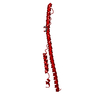
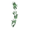
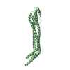
| Deposited unit | 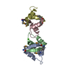
| ||||||||
|---|---|---|---|---|---|---|---|---|---|
| 1 | 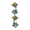
| ||||||||
| 2 | 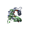
| ||||||||
| 3 | 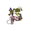
| ||||||||
| Unit cell |
| ||||||||
| Components on special symmetry positions |
| ||||||||
| Noncrystallographic symmetry (NCS) | NCS oper: (Code: given Matrix: (0.2795, 0.4208, -0.863), Vector: Details | Structural Assembly: dimer. Two monomers are related by a dyad axis. The a.s.u. contains two of these dimers (AB and CD molecules) / Biological Assembly: filament. Adjacent dimers are related by a dyad axis (perpendicular to the filament) and a four-fold screw axis (parallel to the filament axis) | |
-Components
| #1: Protein/peptide | Mass: 5695.182 Da / Num. of mol.: 4 / Fragment: N-terminal domain Source method: isolated from a genetically manipulated source Source: (gene. exp.) #2: Chemical | ChemComp-CA / |   Mass: 40.078 Da / Num. of mol.: 1 / Source method: obtained synthetically / Formula: Ca Mass: 40.078 Da / Num. of mol.: 1 / Source method: obtained synthetically / Formula: Ca#3: Water | ChemComp-HOH / |  Mass: 18.015 Da / Num. of mol.: 175 / Source method: isolated from a natural source / Formula: H2O Mass: 18.015 Da / Num. of mol.: 175 / Source method: isolated from a natural source / Formula: H2OHas protein modification | Y | |
|---|
-Experimental details
-Experiment
| Experiment | Method: X-RAY DIFFRACTION / Number of used crystals: 1 |
|---|
- Sample preparation
| Crystal | Density Matthews: 5.04 Å3/Da / Density % sol: 75.39 % |
|---|---|
| Crystal grow | Temperature: 277 K / Method: vapor diffusion, sitting drop / pH: 8 Details: MPD, calcium chloride, TRIS, isopropanol, pH 8, VAPOR DIFFUSION, SITTING DROP, temperature 277K |
-Data collection
| Diffraction |
| ||||||||||||||||||
|---|---|---|---|---|---|---|---|---|---|---|---|---|---|---|---|---|---|---|---|
| Diffraction source |
| ||||||||||||||||||
| Detector |
| ||||||||||||||||||
| Radiation |
| ||||||||||||||||||
| Radiation wavelength |
| ||||||||||||||||||
| Reflection | Resolution: 2.15→25 Å / Num. all: 24769 / Num. obs: 24398 / % possible obs: 98.5 % / Observed criterion σ(F): 0 / Observed criterion σ(I): 0 / Redundancy: 15.7 % / Biso Wilson estimate: 35.4 Å2 / Rmerge(I) obs: 0.076 / Rsym value: 0.054 / Net I/σ(I): 26.1 | ||||||||||||||||||
| Reflection shell | Resolution: 2.15→2.19 Å / Redundancy: 9.8 % / Rmerge(I) obs: 0.46 / Mean I/σ(I) obs: 2.1 / Num. unique all: 1200 / Rsym value: 0.411 / % possible all: 84.6 |

- Processing
| Software |
| ||||||||||||||||||||||||||||||||||||
|---|---|---|---|---|---|---|---|---|---|---|---|---|---|---|---|---|---|---|---|---|---|---|---|---|---|---|---|---|---|---|---|---|---|---|---|---|---|
| Refinement | Method to determine structure: MAD / Resolution: 2.15→24.71 Å / Rfactor Rfree error: 0.006 / Data cutoff high absF: 1242499.83 / Data cutoff low absF: 0 / Isotropic thermal model: RESTRAINED / Cross valid method: THROUGHOUT / σ(F): 0 / σ(I): 0 / Stereochemistry target values: Engh & Huber
| ||||||||||||||||||||||||||||||||||||
| Solvent computation | Solvent model: FLAT MODEL / Bsol: 59.1499 Å2 / ksol: 0.346944 e/Å3 | ||||||||||||||||||||||||||||||||||||
| Displacement parameters | Biso mean: 48.3 Å2
| ||||||||||||||||||||||||||||||||||||
| Refine analyze |
| ||||||||||||||||||||||||||||||||||||
| Refinement step | Cycle: LAST / Resolution: 2.15→24.71 Å
| ||||||||||||||||||||||||||||||||||||
| Refine LS restraints |
| ||||||||||||||||||||||||||||||||||||
| LS refinement shell | Resolution: 2.15→2.28 Å / Rfactor Rfree error: 0.03 / Total num. of bins used: 6
| ||||||||||||||||||||||||||||||||||||
| Xplor file |
|
