 Movie
Movie Controller

[English] 日本語
 Yorodumi
- PDB-1xru: Crystal Structure of 5-keto-4-deoxyuronate Isomerase from Escheri... -
+ Open data

- Basic information
| Entry | Database: PDB / ID: 1xru | ||||||
|---|---|---|---|---|---|---|---|
| Title | Crystal Structure of 5-keto-4-deoxyuronate Isomerase from Eschericia coli | ||||||
 Components Components | 4-deoxy-L-threo-5-hexosulose-uronate ketol-isomerase | ||||||
 Keywords Keywords | ISOMERASE / BETA BARREL / CUPIN | ||||||
| Function / homology |  Function and homology information Function and homology information5-dehydro-4-deoxy-D-glucuronate isomerase / 4-deoxy-L-threo-5-hexosulose-uronate ketol-isomerase activity / D-galacturonate catabolic process / D-glucuronate catabolic process / glucose-6-phosphate isomerase activity / pectin catabolic process / protein homodimerization activity / zinc ion binding / metal ion binding / identical protein binding / cytosol Similarity search - Function | ||||||
| Biological species |  | ||||||
| Method |  X-RAY DIFFRACTION / SYNCHROTRON / MAD / Resolution: 1.94 Å X-RAY DIFFRACTION / SYNCHROTRON / MAD / Resolution: 1.94 Å | ||||||
 Authors Authors | Crowther, R.L. / Georgiadis, M.M. | ||||||
 Citation Citation | Journal: Proteins / Year: 2005 Title: The crystal structure of 5-keto-4-deoxyuronate isomerase from Escherichia coli Authors: Crowther, R.L. / Georgiadis, M.M. | ||||||
| History |
| ||||||
| Remark 999 | SEQUENCE Author states that the C-terminal differences from database sequence is a neutral ...SEQUENCE Author states that the C-terminal differences from database sequence is a neutral designation. The C-terminal sequence reported here is indeed correct for the gene that was cloned and for protein that was crystallized. |
- Structure visualization
| Structure viewer | Molecule: MolmilJmol/JSmol |
|---|
- Downloads & links
-Download
| PDBx/mmCIF format | 1xru.cif.gz | 134.7 KB | Display | PDBx/mmCIF format |
|---|---|---|---|---|
| PDB format | pdb1xru.ent.gz | 104.8 KB | Display | PDB format |
| PDBx/mmJSON format | 1xru.json.gz | Tree view | PDBx/mmJSON format | |
| Others |  Other downloads Other downloads |
-Validation report
| Arichive directory | https://data.pdbj.org/pub/pdb/validation_reports/xr/1xruftp://data.pdbj.org/pub/pdb/validation_reports/xr/1xru | HTTPS FTP |
|---|
-Related structure data
| Similar structure data |
|---|


-Links
 PDBj
PDBj- Assembly
| Deposited unit | 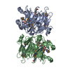
| ||||||||
|---|---|---|---|---|---|---|---|---|---|
| 1 | 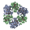
| ||||||||
| 2 | 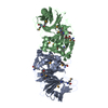
| ||||||||
| Unit cell |
| ||||||||
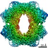
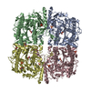
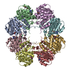
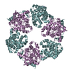
| Details | The biological assembly is a hexamer generated from the dimer in the asymmetric unit by the operations: -y, x-y, z and y-x, -x, z. |
-Components
| #1: Protein | Mass: 31874.428 Da / Num. of mol.: 2 Source method: isolated from a genetically manipulated source Source: (gene. exp.) References: UniProt: Q46938, 5-dehydro-4-deoxy-D-glucuronate isomerase #2: Chemical |   Mass: 65.409 Da / Num. of mol.: 2 / Source method: obtained synthetically / Formula: Zn Mass: 65.409 Da / Num. of mol.: 2 / Source method: obtained synthetically / Formula: Zn#3: Chemical |   Mass: 238.278 Da / Num. of mol.: 2 / Source method: obtained synthetically / Formula: C10H22O6 / Comment: precipitant*YM Mass: 238.278 Da / Num. of mol.: 2 / Source method: obtained synthetically / Formula: C10H22O6 / Comment: precipitant*YM#4: Water | ChemComp-HOH / |  Mass: 18.015 Da / Num. of mol.: 457 / Source method: isolated from a natural source / Formula: H2O Mass: 18.015 Da / Num. of mol.: 457 / Source method: isolated from a natural source / Formula: H2OHas protein modification | Y | |
|---|
-Experimental details
-Experiment
| Experiment | Method: X-RAY DIFFRACTION / Number of used crystals: 1 |
|---|
- Sample preparation
| Crystal | Density Matthews: 2.9 Å3/Da / Density % sol: 56 % |
|---|---|
| Crystal grow | Temperature: 277 K / Method: vapor diffusion / pH: 7 Details: PEG 400, calcium chloride, HEPES, pH 7, VAPOR DIFFUSION, temperature 277K |
-Data collection
| Diffraction | Mean temperature: 100 K | ||||||||||||
|---|---|---|---|---|---|---|---|---|---|---|---|---|---|
| Diffraction source | Source: SYNCHROTRON / Site: NSLS  / Beamline: X8C / Wavelength: 0.980122, 0.979634, 0.979538 / Beamline: X8C / Wavelength: 0.980122, 0.979634, 0.979538 | ||||||||||||
| Detector | Type: ADSC QUANTUM 4 / Detector: CCD / Date: Apr 15, 2002 | ||||||||||||
| Radiation | Monochromator: SAGITALLY FOCUSED Si(111) / Protocol: MAD / Monochromatic (M) / Laue (L): M / Scattering type: x-ray | ||||||||||||
| Radiation wavelength |
| ||||||||||||
| Reflection | Resolution: 1.94→50 Å / Num. all: 51624 / Num. obs: 51573 / % possible obs: 99.9 % / Observed criterion σ(F): 2 / Observed criterion σ(I): 1 / Redundancy: 4.5 % / Rsym value: 0.07 / Net I/σ(I): 9 | ||||||||||||
| Reflection shell | Resolution: 1.94→2.01 Å / Redundancy: 4.5 % / Mean I/σ(I) obs: 4.8 / Num. unique all: 5124 / Rsym value: 0.2 / % possible all: 99.3 |
- Processing
| Software |
| |||||||||||||||||||||||||
|---|---|---|---|---|---|---|---|---|---|---|---|---|---|---|---|---|---|---|---|---|---|---|---|---|---|---|
| Refinement | Method to determine structure: MAD / Resolution: 1.94→12.97 Å / Isotropic thermal model: OVERALL / σ(F): 0 / Stereochemistry target values: Engh & Huber
| |||||||||||||||||||||||||
| Displacement parameters | Biso mean: 16.6 Å2
| |||||||||||||||||||||||||
| Refine analyze | Luzzati coordinate error obs: 0.17 Å / Luzzati d res low obs: 5 Å / Luzzati sigma a obs: 0.2 Å | |||||||||||||||||||||||||
| Refinement step | Cycle: LAST / Resolution: 1.94→12.97 Å
| |||||||||||||||||||||||||
| Refine LS restraints |
|
