 ムービー
ムービー コントローラー


+ データを開く

- 基本情報
基本情報
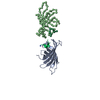
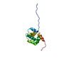
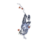
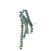
| 登録情報 | データベース: PDB / ID: 1xr0 | ||||||
|---|---|---|---|---|---|---|---|
| タイトル | Structural Basis of SNT PTB Domain Interactions with Distinct Neurotrophic Receptors | ||||||
 要素 要素 |
| ||||||
 キーワード キーワード | SIGNALING PROTEIN/GROWTH FACTOR RECEPTOR / FGFR / SNT / phosphotyrosine binding domain / PTB / TRK / NPXpY motif / SIGNALING PROTEIN-GROWTH FACTOR RECEPTOR COMPLEX | ||||||
| 機能・相同性 |  機能・相同性情報 機能・相同性情報lens placode formation involved in camera-type eye formation / cell surface receptor protein tyrosine phosphatase signaling pathway / negative regulation of cardiac muscle cell differentiation / lens fiber cell development / Signaling by FGFR1 amplification mutants / negative regulation of fibroblast growth factor production / positive regulation of mitotic cell cycle DNA replication / regulation of extrinsic apoptotic signaling pathway in absence of ligand / Signaling by plasma membrane FGFR1 fusions / diphosphate metabolic process ...lens placode formation involved in camera-type eye formation / cell surface receptor protein tyrosine phosphatase signaling pathway / negative regulation of cardiac muscle cell differentiation / lens fiber cell development / Signaling by FGFR1 amplification mutants / negative regulation of fibroblast growth factor production / positive regulation of mitotic cell cycle DNA replication / regulation of extrinsic apoptotic signaling pathway in absence of ligand / Signaling by plasma membrane FGFR1 fusions / diphosphate metabolic process / anterior/posterior axis specification, embryo / regulation of phosphate transport / FGFR1c and Klotho ligand binding and activation / regulation of lateral mesodermal cell fate specification / positive regulation of MAPKKK cascade by fibroblast growth factor receptor signaling pathway / cementum mineralization / vitamin D3 metabolic process / regulation of branching involved in salivary gland morphogenesis by mesenchymal-epithelial signaling / response to sodium phosphate / fibroblast growth factor receptor signaling pathway involved in orbitofrontal cortex development / ventricular zone neuroblast division / receptor-receptor interaction / Epithelial-Mesenchymal Transition (EMT) during gastrulation / positive regulation of phospholipase activity / chordate embryonic development / auditory receptor cell development / positive regulation of parathyroid hormone secretion / mesenchymal cell proliferation / fibroblast growth factor receptor binding / phosphatase activator activity / paraxial mesoderm development / FGFR1b ligand binding and activation / regulation of postsynaptic density assembly / Signaling by activated point mutants of FGFR1 / fibroblast growth factor receptor activity / FGFR1c ligand binding and activation / organ induction / Downstream signaling of activated FGFR1 / neurotrophin TRKA receptor binding / branching involved in salivary gland morphogenesis / Phospholipase C-mediated cascade: FGFR1 / lung-associated mesenchyme development / cell projection assembly / transmembrane receptor protein tyrosine kinase adaptor activity / regulation of epithelial cell proliferation / gastrulation with mouth forming second / outer ear morphogenesis / embryonic limb morphogenesis / positive regulation of endothelial cell chemotaxis / RND1 GTPase cycle / positive regulation of vascular endothelial cell proliferation / RND2 GTPase cycle / positive regulation of mesenchymal cell proliferation / ureteric bud development / middle ear morphogenesis / skeletal system morphogenesis / ventricular septum development / Signaling by ALK / inner ear morphogenesis / PI-3K cascade:FGFR3 / Frs2-mediated activation / PI-3K cascade:FGFR2 / PI-3K cascade:FGFR4 / phosphatidylinositol-mediated signaling / PI-3K cascade:FGFR1 / Formation of paraxial mesoderm / positive regulation of stem cell proliferation / prostate epithelial cord arborization involved in prostate glandular acinus morphogenesis / midbrain development / fibroblast growth factor binding / forebrain development / positive regulation of MAP kinase activity / regulation of cell differentiation / RET signaling / PI3K Cascade / epithelial to mesenchymal transition / fibroblast growth factor receptor signaling pathway / positive regulation of blood vessel endothelial cell migration / neuroblast proliferation / chondrocyte differentiation / cardiac muscle cell proliferation / Activated NTRK2 signals through FRS2 and FRS3 / regulation of ERK1 and ERK2 cascade / calcium ion homeostasis / Signaling by FGFR4 in disease / SHC-mediated cascade:FGFR1 / cell maturation / positive regulation of cardiac muscle cell proliferation / FRS-mediated FGFR3 signaling / FRS-mediated FGFR2 signaling / FRS-mediated FGFR4 signaling / FRS-mediated FGFR1 signaling / Signaling by FGFR3 in disease / cellular response to fibroblast growth factor stimulus / Signaling by FGFR2 in disease / positive regulation of neuron differentiation / Signaling by FGFR1 in disease / NCAM signaling for neurite out-growth / endomembrane system / SH2 domain binding 類似検索 - 分子機能 | ||||||
| 生物種 |  Homo sapiens (ヒト) Homo sapiens (ヒト) | ||||||
| 手法 | 溶液NMR / Structures of the SNT-1 PTB domain in complex with the hFGFR1 peptide were calculated with a distance geometry, simulated annealing protocol by using the X-PLOR program ([4]). NOE distance, dihedral angle restraints were treated with a square-well potential of 50 kcal mol?1 ?2. A total of 2448 manually assigned NOE-derived distance restraints were obtained from the 15N- or 13C-edited NOESY data. Included in this figure are 251 intrapeptide, 258 intermolecular distance restraints. Additionally, 255 unambiguous, 52 ambiguous distance restraints were identified from the NOE data by using ARIA. The final structure calculations employed a total of 2755 NOE restraints obtained from the manual, the ARIA-assisted assignments, 2703 of which were unambiguously assigned NOE-derived distance restraints that comprise 1072 intraresidue, 466 sequential, 216 medium-range, and 949 long-range NOEs. In addition, 70 hydrogen-bond distance restraints for 35 hydrogen bonds, 19 -angle restraints were also used in the structure calculations. For the ensemble of the final 20 structures, no distance or torsional angle restraint was violated by more than 0.4 or 5, respectively. The distance-violation, dihedral-violation, and total energies were 74.4 1.7 kcal mol, 1, 0.82 0.08 kcal mol, 1, and 262.0 6.0 kcal mol, 1, respectively. The Lennard-Jones potential, which was not used during any refinement stage, was 659.3 23.1 kcal mol, 1 for the final structures. Ramachandran plot analysis by Procheck-NMR showed that in the final structures of the complex, 98.1% of the backbone geometries of the non-Gly, non-Pro residues in the complex (protein residues 18-116, peptide residues (412-430), nearly 100% in the secondary structure (protein residues 19-24, 35-40, 45-49, 52-57, 63-68, 71-76, 85-90, 94-107, and 111-115, peptide residues 426-430) lie within energetically favorable or allowed regions. | ||||||
 データ登録者 データ登録者 | Dhalluin, C. / Yan, K.S. / Plotnikova, O. / Lee, K.W. / Zeng, L. / Kuti, M. / Mujtaba, S. / Goldfarb, M.P. / Zhou, M.-M. | ||||||
 引用 引用 | ジャーナル: Mol.Cell / 年: 2000 タイトル: Structural Basis of SNT PTB Domain Interactions with Distinct Neurotrophic Receptors 著者: Dhalluin, C. / Yan, K.S. / Plotnikova, O. / Lee, K.W. / Zeng, L. / Kuti, M. / Mujtaba, S. / Goldfarb, M.P. / Zhou, M.-M. | ||||||
| 履歴 |
|
- 構造の表示
構造の表示
| 構造ビューア | 分子: MolmilJmol/JSmol |
|---|
- ダウンロードとリンク
-ダウンロード
| PDBx/mmCIF形式 | 1xr0.cif.gz | 61.3 KB | 表示 | PDBx/mmCIF形式 |
|---|---|---|---|---|
| PDB形式 | pdb1xr0.ent.gz | 44.9 KB | 表示 | PDB形式 |
| PDBx/mmJSON形式 | 1xr0.json.gz | ツリー表示 | PDBx/mmJSON形式 | |
| その他 |  その他のダウンロード その他のダウンロード |
-検証レポート
| 文書・要旨 | 1xr0_validation.pdf.gz | 256.8 KB | 表示 | wwPDB検証レポート |
|---|---|---|---|---|
| 文書・詳細版 | 1xr0_full_validation.pdf.gz | 256.6 KB | 表示 | |
| XML形式データ | 1xr0_validation.xml.gz | 5.6 KB | 表示 | |
| CIF形式データ | 1xr0_validation.cif.gz | 7 KB | 表示 | |
| アーカイブディレクトリ | https://data.pdbj.org/pub/pdb/validation_reports/xr/1xr0ftp://data.pdbj.org/pub/pdb/validation_reports/xr/1xr0 | HTTPS FTP |
-関連構造データ


-リンク
 PDBj
PDBj


- 集合体
集合体
| 登録構造単位 | 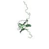
| |||||||||
|---|---|---|---|---|---|---|---|---|---|---|
| 1 |
| |||||||||
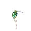
| NMR アンサンブル |
|
-要素
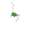
| #1: タンパク質・ペプチド | 分子量: 2492.983 Da / 分子数: 1 断片: Sequence database residues 409-430 from the juxtamembrane region of hFGFR1 由来タイプ: 合成 詳細: The peptide was chemically synthesized. The sequence is taken from Homo sapiens (human). 参照: UniProt: P11362 |
|---|---|
| #2: タンパク質 | 分子量: 15005.791 Da / 分子数: 1 / 断片: PTB domain at the N terminus / 由来タイプ: 組換発現 / 由来: (組換発現) Homo sapiens (ヒト) / 発現宿主:  |
-実験情報
-実験
| 実験 | 手法: 溶液NMR | ||||||||||||||||||||
|---|---|---|---|---|---|---|---|---|---|---|---|---|---|---|---|---|---|---|---|---|---|
| NMR実験 |
| ||||||||||||||||||||
| NMR実験の詳細 | Text: NMR spectra were acquired at 30C on a Bruker DRX600 or DRX500 spectrometer. The backbone and side chain 1H, 13C, and 15N resonances of the protein were assigned using deuterium-decoupled triple- ...Text: NMR spectra were acquired at 30C on a Bruker DRX600 or DRX500 spectrometer. The backbone and side chain 1H, 13C, and 15N resonances of the protein were assigned using deuterium-decoupled triple-resonance experiments of HNCA, HN(CO)CA, HNCACB, HN(CO)CACB, and (H)C(CO)NH-TOCSY ([34 and 30]) recorded by using uniformly 15N/13C-labeled and fractionally deuterated protein in complex with a nonisotopically labeled hFGFR1 peptide. The side chain assignments were completed using 3D HCCH-TOCSY ([7]) data collected from a uniformly 15N/13C labeled-protein/nonlabeled-peptide complex. NOE-derived distance restraints were obtained from 15N- or 13C-edited 3D NOESY spectra ([7]). -angle restraints were determined from 3JHN,H coupling constants measured in a 3D HNHA-J spectrum ([7]). Slowly exchanging amide protons were identified from a series of 2D 15N-HSQC spectra recorded after the H2O buffer was changed to 2H2O buffer. The peptide resonances were assigned using 13C/15N-filtered 2D NOESY and TOCSY spectra ([30]) collected from a 15N/13C labeled-protein/nonlabeled-peptide complex. The intermolecular NOEs used in defining the structure of the SNT-1 PTB domain/hFGFR1 complex were detected in 13C- or 15N-edited (F 1), 13C/15N-filtered (F 3) 3D NOESY spectra. All NMR spectra were processed with NMRPipe/NMRDraw ([8]) and analyzed using NMRView. |
- 試料調製
試料調製
| 詳細 | 内容: SNT-1 PTB domain/hFGFR1 peptide complex (1:1) of ~0.5 mM in 100 mM phosphate buffer of pH 6.5, 5 mM DTT-d10, and 0.5 mM EDTA in H2O/2H2O (9/1) or 2H2O 溶媒系: H2O/2H2O (9/1) or 100% 2H2O |
|---|---|
| 試料状態 | イオン強度: 15 mM DTT-d10, and 0.5 mM EDTA00 mM phosphate buffer, pH: 6.5 / 圧: 1 atm / 温度: 303 K |
-NMR測定
| 放射 | プロトコル: SINGLE WAVELENGTH / 単色(M)・ラウエ(L): M | |||||||||||||||
|---|---|---|---|---|---|---|---|---|---|---|---|---|---|---|---|---|
| 放射波長 | 相対比: 1 | |||||||||||||||
| NMRスペクトロメーター |
|
- 解析
解析
| NMR software |
| ||||||||||||
|---|---|---|---|---|---|---|---|---|---|---|---|---|---|
| 精密化 | 手法: Structures of the SNT-1 PTB domain in complex with the hFGFR1 peptide were calculated with a distance geometry, simulated annealing protocol by using the X-PLOR program ([4]). NOE distance, ...手法: Structures of the SNT-1 PTB domain in complex with the hFGFR1 peptide were calculated with a distance geometry, simulated annealing protocol by using the X-PLOR program ([4]). NOE distance, dihedral angle restraints were treated with a square-well potential of 50 kcal mol?1 ?2. A total of 2448 manually assigned NOE-derived distance restraints were obtained from the 15N- or 13C-edited NOESY data. Included in this figure are 251 intrapeptide, 258 intermolecular distance restraints. Additionally, 255 unambiguous, 52 ambiguous distance restraints were identified from the NOE data by using ARIA. The final structure calculations employed a total of 2755 NOE restraints obtained from the manual, the ARIA-assisted assignments, 2703 of which were unambiguously assigned NOE-derived distance restraints that comprise 1072 intraresidue, 466 sequential, 216 medium-range, and 949 long-range NOEs. In addition, 70 hydrogen-bond distance restraints for 35 hydrogen bonds, 19 -angle restraints were also used in the structure calculations. For the ensemble of the final 20 structures, no distance or torsional angle restraint was violated by more than 0.4 or 5, respectively. The distance-violation, dihedral-violation, and total energies were 74.4 1.7 kcal mol, 1, 0.82 0.08 kcal mol, 1, and 262.0 6.0 kcal mol, 1, respectively. The Lennard-Jones potential, which was not used during any refinement stage, was 659.3 23.1 kcal mol, 1 for the final structures. Ramachandran plot analysis by Procheck-NMR showed that in the final structures of the complex, 98.1% of the backbone geometries of the non-Gly, non-Pro residues in the complex (protein residues 18-116, peptide residues (412-430), nearly 100% in the secondary structure (protein residues 19-24, 35-40, 45-49, 52-57, 63-68, 71-76, 85-90, 94-107, and 111-115, peptide residues 426-430) lie within energetically favorable or allowed regions. ソフトェア番号: 1 | ||||||||||||
| 代表構造 | 選択基準: fewest violations | ||||||||||||
| NMRアンサンブル | コンフォーマー選択の基準: structures with the least restraint violations 計算したコンフォーマーの数: 100 / 登録したコンフォーマーの数: 1 |
 X-PLOR