 Movie
Movie Controller

[English] 日本語
 Yorodumi
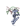
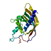
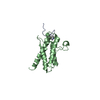
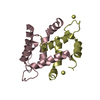
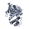
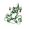
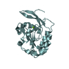
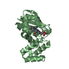
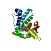
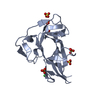
- PDB-1xok: crystal structure of alfalfa mosaic virus RNA 3'UTR in complex wi... -
+ Open data

- Basic information
| Entry | Database: PDB / ID: 1xok | ||||||
|---|---|---|---|---|---|---|---|
| Title | crystal structure of alfalfa mosaic virus RNA 3'UTR in complex with coat protein N terminal peptide | ||||||
 Components Components |
| ||||||
 Keywords Keywords | Viral protein/RNA / protein-rna complex / alpha helix / atypical rna duplex / Viral protein-RNA COMPLEX | ||||||
| Function / homology | T=3 icosahedral viral capsid / viral nucleocapsid / ribonucleoprotein complex / RNA binding / BROMIDE ION / RNA / RNA (> 10) / Capsid protein Function and homology information Function and homology information | ||||||
| Method |  X-RAY DIFFRACTION / SYNCHROTRON / MAD / Resolution: 3 Å X-RAY DIFFRACTION / SYNCHROTRON / MAD / Resolution: 3 Å | ||||||
 Authors Authors | Guogas, L.M. / Filman, D.J. / Hogle, J.M. / Gehrke, L. | ||||||
 Citation Citation | Journal: Science / Year: 2004 Title: Cofolding organizes alfalfa mosaic virus RNA and coat protein for replication. Authors: Guogas, L.M. / Filman, D.J. / Hogle, J.M. / Gehrke, L. | ||||||
| History |
|
- Structure visualization
| Structure viewer | Molecule: MolmilJmol/JSmol |
|---|
- Downloads & links
-Download
| PDBx/mmCIF format | 1xok.cif.gz | 38.6 KB | Display | PDBx/mmCIF format |
|---|---|---|---|---|
| PDB format | pdb1xok.ent.gz | 26.3 KB | Display | PDB format |
| PDBx/mmJSON format | 1xok.json.gz | Tree view | PDBx/mmJSON format | |
| Others |  Other downloads Other downloads |
-Validation report
| Arichive directory | https://data.pdbj.org/pub/pdb/validation_reports/xo/1xokftp://data.pdbj.org/pub/pdb/validation_reports/xo/1xok | HTTPS FTP |
|---|
-Related structure data
| Similar structure data |
|---|
-Links
 PDBj
PDBj


- Assembly
| Deposited unit | 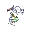
| ||||||||
|---|---|---|---|---|---|---|---|---|---|
| 1 |
| ||||||||
| Unit cell |
|
-Components
| #1: RNA chain | Mass: 9533.723 Da / Num. of mol.: 1 / Fragment: amv rna bases 843-872 / Source method: obtained synthetically / Details: rna synthesis dharmacon research | ||||
|---|---|---|---|---|---|
| #2: RNA chain | Mass: 2934.831 Da / Num. of mol.: 1 / Fragment: amv rna bases 873-881 / Source method: obtained synthetically / Details: rna synthesis dharmacon research | ||||
| #3: Protein/peptide | Mass: 2756.170 Da / Num. of mol.: 2 / Fragment: amv coat protein residues 1-26 / Source method: obtained synthetically / Details: peptide synthesis by mit biopolymers laboratory / References: UniProt: P24264 #4: Chemical | ChemComp-BR / |   Mass: 79.904 Da / Num. of mol.: 1 / Source method: obtained synthetically / Formula: Br Mass: 79.904 Da / Num. of mol.: 1 / Source method: obtained synthetically / Formula: Br#5: Water | ChemComp-HOH / |  Mass: 18.015 Da / Num. of mol.: 7 / Source method: isolated from a natural source / Formula: H2O Mass: 18.015 Da / Num. of mol.: 7 / Source method: isolated from a natural source / Formula: H2O |
-Experimental details
-Experiment
| Experiment | Method: X-RAY DIFFRACTION / Number of used crystals: 2 |
|---|
- Sample preparation
| Crystal | Density Matthews: 2.3 Å3/Da / Density % sol: 46 % |
|---|---|
| Crystal grow | Temperature: 295 K / Method: vapor diffusion, hanging drop / pH: 5.6 Details: 2.45M ammonium sulfate, 0.01M magnesium acetate, 0.01M mes, 0.001M samarium chloride, pH 5.6, VAPOR DIFFUSION, HANGING DROP, temperature 295K |
-Data collection
| Diffraction |
| ||||||||||||||||||
|---|---|---|---|---|---|---|---|---|---|---|---|---|---|---|---|---|---|---|---|
| Diffraction source |
| ||||||||||||||||||
| Detector |
| ||||||||||||||||||
| Radiation |
| ||||||||||||||||||
| Radiation wavelength |
| ||||||||||||||||||
| Reflection | Resolution: 3→99 Å / Num. all: 3508 / Num. obs: 3487 / % possible obs: 99.4 % / Observed criterion σ(F): 0 / Observed criterion σ(I): 0 / Redundancy: 11.9 % / Rsym value: 0.085 / Net I/σ(I): 25 | ||||||||||||||||||
| Reflection shell | Resolution: 3→3.19 Å / Mean I/σ(I) obs: 4.4 / Rsym value: 0.405 / % possible all: 98.1 |

- Processing
| Software |
| ||||||||||||||||||||||||||||||||||||||||||||||||||||||||||||||||||||||
|---|---|---|---|---|---|---|---|---|---|---|---|---|---|---|---|---|---|---|---|---|---|---|---|---|---|---|---|---|---|---|---|---|---|---|---|---|---|---|---|---|---|---|---|---|---|---|---|---|---|---|---|---|---|---|---|---|---|---|---|---|---|---|---|---|---|---|---|---|---|---|---|
| Refinement | Method to determine structure: MAD / Resolution: 3→20 Å / Cor.coef. Fo:Fc: 0.91 / Cor.coef. Fo:Fc free: 0.903 / SU B: 18.921 / SU ML: 0.332 / TLS residual ADP flag: LIKELY RESIDUAL / Cross valid method: THROUGHOUT / σ(F): 0.001 / ESU R Free: 0.467 Stereochemistry target values: MAXIMUM LIKELIHOOD WITH PHASES
| ||||||||||||||||||||||||||||||||||||||||||||||||||||||||||||||||||||||
| Solvent computation | Ion probe radii: 0.8 Å / Shrinkage radii: 0.8 Å / VDW probe radii: 1.4 Å / Solvent model: BABINET MODEL WITH MASK | ||||||||||||||||||||||||||||||||||||||||||||||||||||||||||||||||||||||
| Displacement parameters | Biso mean: 45.218 Å2
| ||||||||||||||||||||||||||||||||||||||||||||||||||||||||||||||||||||||
| Refinement step | Cycle: LAST / Resolution: 3→20 Å
| ||||||||||||||||||||||||||||||||||||||||||||||||||||||||||||||||||||||
| Refine LS restraints |
| ||||||||||||||||||||||||||||||||||||||||||||||||||||||||||||||||||||||
| LS refinement shell | Highest resolution: 3 Å / Num. reflection Rwork: 453 / Total num. of bins used: 10 | ||||||||||||||||||||||||||||||||||||||||||||||||||||||||||||||||||||||
| Refinement TLS params. | Method: refined / Origin x: 36.6597 Å / Origin y: 13.0441 Å / Origin z: 22.9456 Å
| ||||||||||||||||||||||||||||||||||||||||||||||||||||||||||||||||||||||
| Refinement TLS group |
|
