- PDB-1xnn: CRYSTAL STRUCTURE OF THE RAT ANDROGEN RECEPTOR LIGAND BINDING DOM... -
+
Open data
ID or keywords:
Loading...
-
Basic information
Entry
Database: PDB / ID: 1xnn
Title
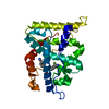
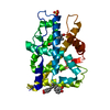
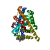
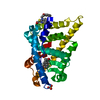
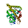
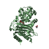
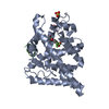
CRYSTAL STRUCTURE OF THE RAT ANDROGEN RECEPTOR LIGAND BINDING DOMAIN T877A MUTANT COMPLEX WITH (3A-ALPHA-,4-ALPHA 7-ALPHA-,7A-ALPHA-)-3A,4,7,7A-TETRAHYDRO-2-(4-NITRO-1-NAPHTHALENYL)-4,7-ETHANO-1H-ISOINDOLE-1,3(2H)-DIONE.
HELIX DETERMINATION METHOD: PROCHECK UNDER STANDARD NUCLEAR RECEPTOR LIGAND BINDING DOMAIN ... HELIX DETERMINATION METHOD: PROCHECK UNDER STANDARD NUCLEAR RECEPTOR LIGAND BINDING DOMAIN NUMBERING, HELIX 2 IS ABSENT AND HELICES 10 AND 11 ARE CONTIGUOUS IN THIS STRUCTURE.
Remark 700
SHEET DETERMINATION METHOD: AUTHOR PROVIDED
Remark 999
SEQUENCE HUMAN NUMBERING SCHEME USED. THE CLONE CONTAINED BOTH A HIS-TAG AND A THROMBIN CLEAVAGE ... SEQUENCE HUMAN NUMBERING SCHEME USED. THE CLONE CONTAINED BOTH A HIS-TAG AND A THROMBIN CLEAVAGE SITE. THE PROTEIN IN THE CRYSTAL CONTAINED THE SEQUENCE G S H M FOLLOWED BY THE PROTEINS RESIDUES 664 TO 919.
In the structure databanks used in Yorodumi, some data are registered as the other names, "COVID-19 virus" and "2019-nCoV". Here are the details of the virus and the list of structure data.
Jan 31, 2019. EMDB accession codes are about to change! (news from PDBe EMDB page)
EMDB accession codes are about to change! (news from PDBe EMDB page)
The allocation of 4 digits for EMDB accession codes will soon come to an end. Whilst these codes will remain in use, new EMDB accession codes will include an additional digit and will expand incrementally as the available range of codes is exhausted. The current 4-digit format prefixed with “EMD-” (i.e. EMD-XXXX) will advance to a 5-digit format (i.e. EMD-XXXXX), and so on. It is currently estimated that the 4-digit codes will be depleted around Spring 2019, at which point the 5-digit format will come into force.
The EM Navigator/Yorodumi systems omit the EMD- prefix.
Related info.:Q: What is EMD? / ID/Accession-code notation in Yorodumi/EM Navigator
Yorodumi is a browser for structure data from EMDB, PDB, SASBDB, etc.
This page is also the successor to EM Navigator detail page, and also detail information page/front-end page for Omokage search.
The word "yorodu" (or yorozu) is an old Japanese word meaning "ten thousand". "mi" (miru) is to see.
Related info.:EMDB / PDB / SASBDB / Comparison of 3 databanks / Yorodumi Search / Aug 31, 2016. New EM Navigator & Yorodumi / Yorodumi Papers / Jmol/JSmol / Function and homology information / Changes in new EM Navigator and Yorodumi
 Movie
Movie Controller

 Yorodumi
 Open data
 Basic information
 Components
 Keywords
 Function and homology information
 X-RAY DIFFRACTION /
  Authors
 Citation
 Structure visualization
 Downloads & links
 Other downloads
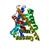


 PDBj
PDBj

 Assembly


 Mass: 350.368 Da / Num. of mol.: 1 / Source method: obtained synthetically / Formula: C20H18N2O4
 Mass: 18.015 Da / Num. of mol.: 35 / Source method: isolated from a natural source / Formula: H2O
 Sample preparation
 / Beamline: 17-ID / Wavelength: 1 / Wavelength: 1 Å
 Processing